Review
. 2015 Aug:33:68-75.
doi: 10.1016/j.sbi.2015.07.012. Epub 2015 Aug 15.

A structural biology perspective on NMDA receptor pharmacology and function

Affiliations
Review

A structural biology perspective on NMDA receptor pharmacology and function

Michael C Regan et al. Curr Opin Struct Biol. 2015 Aug.

Abstract

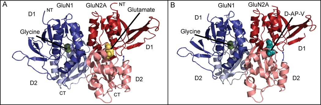
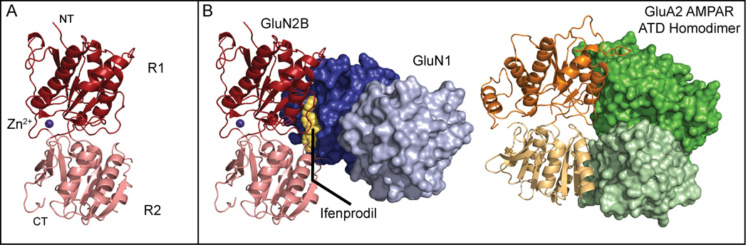
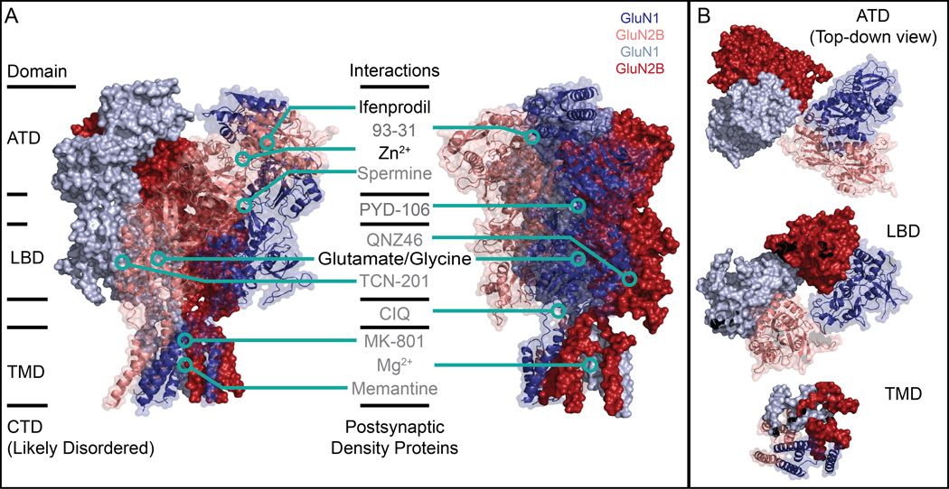
N-methyld-aspartate receptors (NMDARs) belong to the large family of ionotropic glutamate receptors (iGluRs), which are critically involved in basic brain functions as well as multiple neurological diseases and disorders. The NMDARs are large heterotetrameric membrane protein complexes. The extensive extracellular domains recognize neurotransmitter ligands and allosteric compounds and translate the binding information to regulate activity of the transmembrane ion channel. Here, we review recent advances in the structural biology of NMDARs with a focus on pharmacology and function. Structural analysis of the isolated extracellular domains in combination with the intact heterotetrameric NMDAR structure provides important insights into how this sophisticated ligand-gated ion channel may function.


Figures

Figure 1
Figure 1
Structure of the GluN1/ GluN2A LBD heterodimer. GluN1 is shown in blues and GluN2A is shown in reds. A) GluN1 in complex with glycine and GluN2A with glutamate (PDB: 4NF8). The clamshell is in a closed conformation (see arrow between GluN2A upper and lower lobes). B) GluN1 in complex with glycine and GluN2A with AP-V (PDB: 4NF5). Here, the clamshell is in an open state (see arrow between GluN2A upper and lower lobes). The d-AP-V site is shared among all competitive antagonists. Hypothetical binding sites for allosteric antagonists have not been structurally confirmed.
Figure 2
Figure 2
Composite structure and ligand binding of the NMDAR ATD. (A) The isolated GluN2B ATD (PDB: 3JPW) adopts a clamshell-like structure consisting of an upper (R1, dark red) and lower (R2, pink) lobe. Zinc (purple) binds near the hinge between the two lobes and allosterically inhibits GluN2A, and to a lesser extent GluN2B, receptors. (B) Contrary to early work, the allosteric antagonist ifenprodil (yellow) binds between the GluN1b/GluN2B ATD subunits (blues, R1 and R2 lobes, and reds, R1 and R2 lobes, respectively) stabilizing dimer formation (PDB: 3QEL). While the R1 lobes of the GluN1b and GluN2B ATDs are closely apposed, there is practically no interaction between the R2 lobes in the dimer. Conversely, the AMPAR ATD homodimer (PDB: 3H5V) exhibits extensive interactions between both R1 and R2 lobes.
Figure 3
Figure 3
Architecture of the intact NMDAR and selected interactions. (A) The heterotetrameric GluN1a/GluN2B NMDAR (PDB: 4PE5) consists of four domains: the extracellular ATD and LBD, the transmembrane TMD, and the disordered intracellular CTD. A number of small molecule and protein interaction partners have been identified; the structure of the intact receptor provides a framework to visualize where these interactions occur in relation to each other in addition to potential mechanical and pharmacological modulatory mechanisms. Critically, the binding sites of many of these small molecules are as yet putative at best. Several binding sites have been visualized at high-resolution by x-ray crystallography, while for others point mutagenesis studies suggest that the different small molecules bind at distinct sites on the NMDAR. In this figure, small molecules listed in black have been verified by x-ray crystallography while those in grey have not. (B) The domain swap between ATD and LBD is a general feature of iGluRs. In this 1-2-1-2 arrangement, two pairs of GluN1a (blues) /GluN2B (reds) ATD dimers form, shown here as a dark blue/pink dimer and a light blue/dark red dimer. At the LBD, however, the dimers are instead composed of the dark blue/dark red pair and the pink/light blue pair. The TMD shows quasi-fourfold symmetry, with the ion channel pore in the center of this structure in what is believed to be the closed state.

References

    1. Traynelis SF, Wollmuth LP, Mcbain CJ, Menniti FS, Vance KM, Ogden KK, Hansen KB, Yuan H, Myers SJ, Dingledine R. Glutamate receptor ion channels: structure, regulation, and function. Pharmacol. Rev. 2010;62:405–496. - PMC - PubMed
    1. Paoletti P, Bellone C, Zhou Q. NMDA receptor subunit diversity: impact on receptor properties, synaptic plasticity and disease. Nat. Rev. Neurosci. 2013;14:383–400. - PubMed
    1. Zhu S, Paoletti P. Allosteric modulators of NMDA receptors: multiple sites and mechanisms. Curr. Opin. Pharmacol. 2015;20:14–23. - PubMed
    1. Hansen KB, Ogden KK, Yuan H, Traynelis SF. Distinct functional and pharmacological properties of triheteromeric GluN1/GluN2A/GluN2B NMDA receptors. Neuron. 2014;81:1084–1096. - PMC - PubMed
    1. Glasgow NG, Siegler Retchless B, Johnson JW. Molecular bases of NMDA receptor subtype-dependent properties. J. Physiol. 2014;593:83–95. - PMC - PubMed

Publication types