. 2015 Sep 1;112(35):11078-83.
doi: 10.1073/pnas.1507104112. Epub 2015 Aug 17.

Human hippocampus represents space and time during retrieval of real-world memories

Affiliations

Human hippocampus represents space and time during retrieval of real-world memories

Dylan M Nielson et al. Proc Natl Acad Sci U S A. .

Abstract

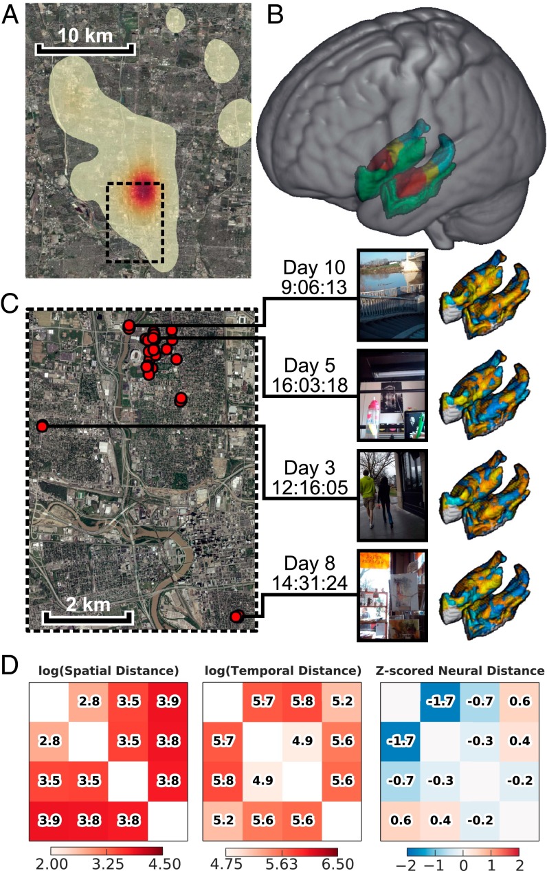
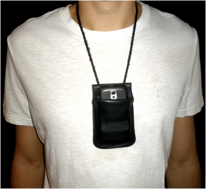
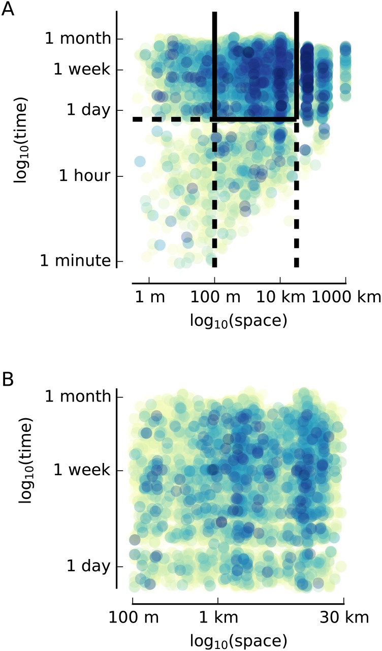
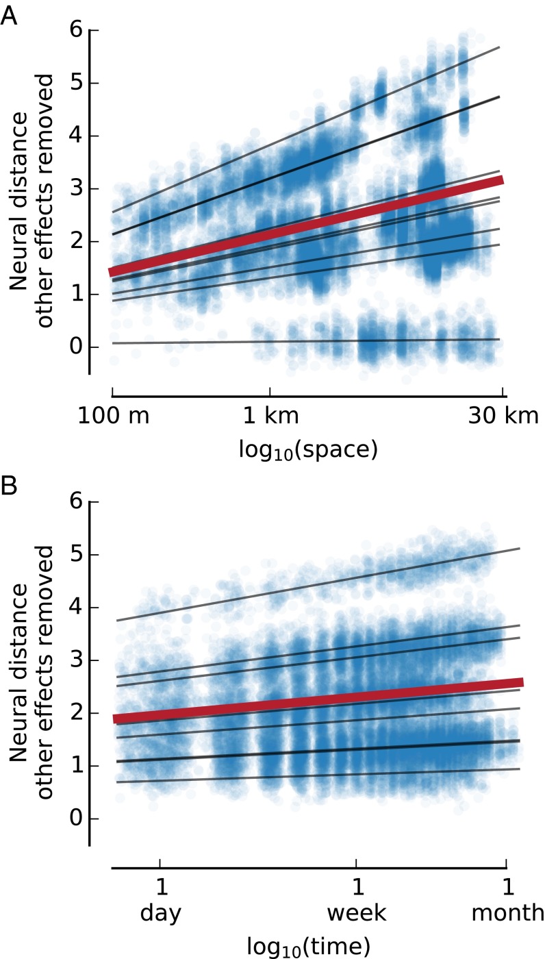
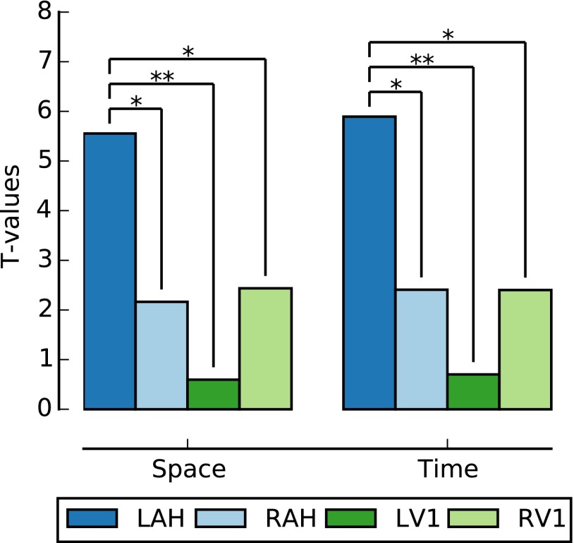
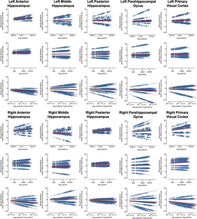
Memory stretches over a lifetime. In controlled laboratory settings, the hippocampus and other medial temporal lobe brain structures have been shown to represent space and time on the scale of meters and seconds. It remains unclear whether the hippocampus also represents space and time over the longer scales necessary for human episodic memory. We recorded neural activity while participants relived their own experiences, cued by photographs taken with a custom lifelogging device. We found that the left anterior hippocampus represents space and time for a month of remembered events occurring over distances of up to 30 km. Although previous studies have identified similar drifts in representational similarity across space or time over the relatively brief time scales (seconds to minutes) that characterize individual episodic memories, our results provide compelling evidence that a similar pattern of spatiotemporal organization also exists for organizing distinct memories that are distant in space and time. These results further support the emerging view that the anterior, as opposed to posterior, hippocampus integrates distinct experiences, thereby providing a scaffold for encoding and retrieval of autobiographical memories on the scale of our lives.

Keywords: episodic memory; hippocampus; lifelogging; representational similarity analysis.


Conflict of interest statement

The authors declare no conflict of interest.

Figures

Fig. 1.
Fig. 1.
Overview of study method. (A) Heat map of locations where images from all subjects were taken. (B) MTL ROIs (red, anterior hippocampus; yellow, middle hippocampus; blue, posterior hippocampus; green, parahippocampus). (C) Subset of the data collected from a single participant. Each red point on the map corresponds to an image shown to the participant in the scanner. Four sample images are displayed, along with the time taken, with the lines indicating the location. The heat maps show the single-trial beta corresponding to each image. (D) Example of the spatial, temporal, and neural distance matrices derived from these four sample images. (Map data courtesy of the US Geological Survey.)
Fig. S1.
Fig. S1.
Example of an Android smartphone in a pouch used for data collection.
Fig. S2.
Fig. S2.
Memory performance. This figure shows the percentage of presented images recalled by each participant. The average recall performance was 63.4%, although performance is not normally distributed across subjects.
Fig. S3.
Fig. S3.
Spatial and temporal distances between pairs of images. (A) Spatial and temporal distances for each pair of images from all subjects. Spatial and temporal distances were only calculated for image pairs if both images were collected by the same subject. The color of the dot indicates the density of points in that region. To be included in subsequent analyses, we required that image pairs have a spatial distance between 100 m and 30 km and a temporal distance greater than 16 h. Image pairs with a distance of 0 m are not shown. (B) Image pairs meeting these criteria are shown.
Fig. 2.
Fig. 2.
Space and time are correlated with neural distance in the left anterior hippocampus. (A and B) Each dot represents a pair of presented images. The black lines show the estimated neural distance based on the regression for each subject. The red line shows the estimated neural distance averaged across subjects. Spatial distance (A) and temporal distance (B) are both correlated with neural distance in the left anterior hippocampus after removing the effects of other factors in the model (i.e., after isolating the effect of space or time).
Fig. 3.
Fig. 3.
Comparison of space and time representations across the brain region. Space and time are significantly more strongly related to neural distance in the left anterior hippocampus (LAH) than in the right anterior hippocampus (RAH) or the right or left primary visual cortex (RV1 and LV1, respectively). *P < 0.05; **P < 0.01.
Fig. S4.
Fig. S4.
Scatter plots for neural distance vs. time, space, and their interaction with other effects removed. The black lines show the predicted neural distance based on the regression for each subject. The red line shows the predicted neural distance averaged across subjects. The left anterior hippocampus is the only region with significant effects (Bonferroni corrected P < 0.01 for space and time, P < 0.05 for their interaction). None of the other regions have a P value less than 0.1 after Bonferroni correction. m + s, meters + seconds.
Fig. S5.
Fig. S5.
Effect of recency on image recall. This figure shows the distribution of times between when images were collected and when they were assessed for recalled and not recalled events for each participant. Participant 2 showed a significant relationship between temporal recency and recall, but the direction of this effect implies weaker memory for more recent events, and it was not strong enough to survive a multiple comparison correction (t = 2.16, uncorrected P = 0.03). Participant 5 recalled the events associated with all of the presented images. Participant 6 is not shown because she dropped out of the study before the fMRI scan was collected.
Fig. S6.
Fig. S6.
Effect of proximity on image recall. This figure shows the distribution of distances between the location where images were collected and the fMRI scanner for recalled and not recalled events for each participant. Participant 7 showed a significant relationship between spatial proximity and recall, but this effect was not strong enough to survive multiple comparison correction (t = −2.54, uncorrected P = 0.01). Participant 5 recalled the events associated with all of the presented images. Participant 6 is not shown because she dropped out of the study before the fMRI scan was collected.
Fig. S7.
Fig. S7.
Contiguity effect of space and time on subsequently recalled images. The first column shows the spatial and temporal distances between pairs of images in which the first item and the second item were both recalled. The second column shows the spatial and temporal distances between pairs of images in which the first item was recalled but the second item was not. The last column plots the distribution of across-subject t values obtained via permutation for the effect of space, time, and their interaction on memory for second items. The true value is indicated by a solid line; dashed lines indicate significance. The first row includes all pairs of images without limiting the spatial or temporal distance between them. There is a clear region between 1 and 100 m where there are more remembered items than not remembered items. This finding is reflected in the significance of space (t = 2.45, P = 0.034), time (t = 2.79, P = 0.022), and their interaction (t = −3.12, P = 0.008). The second row eliminates pairs of images from the same event, and the increased number of remembered vs. not remembered items is no longer present. The last row focuses on the data used in our main analysis, and it is clear that the spatiotemporal distributions of recalled and not recalled items are similar in this region.

References

    1. Scoville WB, Milner B. Loss of recent memory after bilateral hippocampal lesions. J Neurol Neurosurg Psychiatry. 1957;20(1):11–21. - PMC - PubMed
    1. O’Keefe J, Dostrovsky J. The hippocampus as a spatial map. Preliminary evidence from unit activity in the freely-moving rat. Brain Res. 1971;34(1):171–175. - PubMed
    1. O’Keefe J, Burgess N. Geometric determinants of the place fields of hippocampal neurons. Nature. 1996;381(6581):425–428. - PubMed
    1. Hafting T, Fyhn M, Molden S, Moser M-B, Moser EI. Microstructure of a spatial map in the entorhinal cortex. Nature. 2005;436(7052):801–806. - PubMed
    1. McNaughton BL, Battaglia FP, Jensen O, Moser EI, Moser M-B. Path integration and the neural basis of the ‘cognitive map’. Nat Rev Neurosci. 2006;7(8):663–678. - PubMed

Publication types

LinkOut - more resources