. 2015 Sep 1;112(35):10926-31.
doi: 10.1073/pnas.1501430112. Epub 2015 Aug 17.

Viral fusion protein transmembrane domain adopts β-strand structure to facilitate membrane topological changes for virus-cell fusion

Affiliations

Viral fusion protein transmembrane domain adopts β-strand structure to facilitate membrane topological changes for virus-cell fusion

Hongwei Yao et al. Proc Natl Acad Sci U S A. .

Abstract

The C-terminal transmembrane domain (TMD) of viral fusion proteins such as HIV gp41 and influenza hemagglutinin (HA) is traditionally viewed as a passive α-helical anchor of the protein to the virus envelope during its merger with the cell membrane. The conformation, dynamics, and lipid interaction of these fusion protein TMDs have so far eluded high-resolution structure characterization because of their highly hydrophobic nature. Using magic-angle-spinning solid-state NMR spectroscopy, we show that the TMD of the parainfluenza virus 5 (PIV5) fusion protein adopts lipid-dependent conformations and interactions with the membrane and water. In phosphatidylcholine (PC) and phosphatidylglycerol (PG) membranes, the TMD is predominantly α-helical, but in phosphatidylethanolamine (PE) membranes, the TMD changes significantly to the β-strand conformation. Measured order parameters indicate that the strand segments are immobilized and thus oligomerized. (31)P NMR spectra and small-angle X-ray scattering (SAXS) data show that this β-strand-rich conformation converts the PE membrane to a bicontinuous cubic phase, which is rich in negative Gaussian curvature that is characteristic of hemifusion intermediates and fusion pores. (1)H-(31)P 2D correlation spectra and (2)H spectra show that the PE membrane with or without the TMD is much less hydrated than PC and PG membranes, suggesting that the TMD works with the natural dehydration tendency of PE to facilitate membrane merger. These results suggest a new viral-fusion model in which the TMD actively promotes membrane topological changes during fusion using the β-strand as the fusogenic conformation.

Keywords: conformational polymorphism; membrane curvature; peptide–membrane interactions; small-angle X-ray scattering; solid-state NMR spectroscopy.


Conflict of interest statement

The authors declare no conflict of interest.

Figures
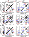
Fig. 1.
Fig. 1.
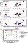
Two-dimensional 13C-13C correlation spectra of the PIV5 TMD in POPC/cholesterol (Left) and POPC (Right) (A), DOPC/DOPG (B), and DOPE membranes (C). (AC) Low-temperature spectra (243–253 K) (Left) allow quantification of the helix and strand intensities, whereas high-temperature spectra (293–303 K) (Right) report the peptide mobility in liquid-crystalline membranes. α-Helix signals (red) dominate in A and B, whereas β-strand signals (blue) become significant in the PE membrane. Superscripts “h” and “s” denote helix and strand, respectively, and ovals highlight the A490 and V506 peaks.
Fig. 2.
Fig. 2.
(AC) Low-temperature 2D 15N-13C correlation spectra of the TMD bound to POPC/POPG (A), DOPC/DOPG (B), and DOPE membranes (C). The TMD is mainly α-helical in the POPC/POPG membrane but becomes more β-strand in the DOPE membrane, consistent with the 2D 13C-13C spectra. (D) Residue-specific helix fractions of membrane-bound TMD obtained from Cα-Cβ or Cα/β-CO cross-peak intensities. The TMD is mostly helical in PC and PG membranes but switches to the strand conformation in PE membranes.
Fig. 3.
Fig. 3.
Two-dimensional 1H-13C correlation spectra of POPE-bound TMD. (A) Two-dimensional spectrum with a 100-ms 1H spin-diffusion mixing time. (B) 1H cross-sections of the sum of β-strand signals as a function of mixing time. (C) Water–peptide (black) and lipid–peptide polarization transfer curves for the β-strand (blue) and α-helical (red) signals. The fast buildup indicates that both helical and strand conformations are well inserted into the membrane.
Fig. 4.
Fig. 4.
Effects of the TMD on membrane curvature and hydration. (A) Static 31P spectra of POPC, POPC/POPG, DOPC/DOPG, and POPE membranes without (black) and with (red) the TMD. (B) Static 31P spectra of DOPE without and with the TMD from 273 K to 303 K. The TMD converted the lamellar and HII powder patterns to an isotropic peak. Deconvolution of the 303 K spectrum shows that ∼15% of the intensity remained in the HII phase (green). (C) Two-dimensional MAS 1H-31P correlation spectra of POPC and POPE membranes with 100-ms 1H spin diffusion. (D and E) 1H cross-sections of the 2D 1H-31P spectra. (D) The PC and PG membranes exhibit a strong water cross-peak. (E) POPE membranes with (black) and without (brown) the TMD show a broad water cross-peak, indicating low hydration.
Fig. 5.
Fig. 5.
The TMD generates NGC to the DOPE membrane. (A) SAXS spectrum of TMD-bound DOPE. An Ia3d cubic phase (red) coexists with an HII phase (green). (Inset) An expanded view of the higher order reflections. (B) Measured Q positions versus assigned reflections in terms of Miller indices h, k, and l. Qmeas is plotted versus (h2+k2+l2) for the cubic phase and (h2+hk+k2) for the HII phase. (C) A catenoid surface with a 6-nm diameter (c = 3 nm) has K = −0.111 nm−2 at its narrowest cross-section. A hemifusion stalk or fusion pore will have a neck that conforms to the catenoid. Arrows indicate directions of negative and positive principal curvatures.
Fig. 6.
Fig. 6.
Structural model of TMD in PE-rich membranes. The TMD adopts a transmembrane strand–helix–strand conformation, which imparts different curvatures to the two leaflets of the membrane. The β-strand segments are immobilized by oligomerization. In these PE-rich membranes, previous data showed that the FP (green) is also rich in β-strand conformation.

References

    1. Lamb RA, Jardetzky TS. Structural basis of viral invasion: Lessons from paramyxovirus F. Curr Opin Struct Biol. 2007;17(4):427–436. - PMC - PubMed
    1. Harrison SC. Viral membrane fusion. Nat Struct Mol Biol. 2008;15(7):690–698. - PMC - PubMed
    1. Eckert DM, Kim PS. Mechanisms of viral membrane fusion and its inhibition. Annu Rev Biochem. 2001;70:777–810. - PubMed
    1. Tamm LK. Hypothesis: Spring-loaded boomerang mechanism of influenza hemagglutinin-mediated membrane fusion. Biochim Biophys Acta. 2003;1614(1):14–23. - PubMed
    1. White JM, Delos SE, Brecher M, Schornberg K. Structures and mechanisms of viral membrane fusion proteins: Multiple variations on a common theme. Crit Rev Biochem Mol Biol. 2008;43(3):189–219. - PMC - PubMed

Publication types

MeSH terms