. 2015 Sep 1;112(35):11084-9.
doi: 10.1073/pnas.1504172112. Epub 2015 Aug 17.

Multiple component networks support working memory in prefrontal cortex

Affiliations

Multiple component networks support working memory in prefrontal cortex

David A Markowitz et al. Proc Natl Acad Sci U S A. .

Erratum in

Abstract

Lateral prefrontal cortex (PFC) is regarded as the hub of the brain's working memory (WM) system, but it remains unclear whether WM is supported by a single distributed network or multiple specialized network components in this region. To investigate this problem, we recorded from neurons in PFC while monkeys made delayed eye movements guided by memory or vision. We show that neuronal responses during these tasks map to three anatomically specific modes of persistent activity. The first two modes encode early and late forms of information storage, whereas the third mode encodes response preparation. Neurons that reflect these modes are concentrated at different anatomical locations in PFC and exhibit distinct patterns of coordinated firing rates and spike timing during WM, consistent with distinct networks. These findings support multiple component models of WM and consequently predict distinct failures that could contribute to neurologic dysfunction.

Keywords: coherence; macaque; prefrontal cortex; working memory.


Conflict of interest statement

The authors declare no conflict of interest.

Figures
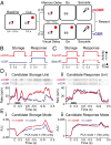
Fig. 1.
Fig. 1.
Experimental design for testing the distinct mode hypothesis. (A) ODR paradigm with interleaved mODR and vODR trials. (B and C) Predicted responses of neurons that encode stored information and prepared responses as a function of (B) delay type and (C) reaction time after a memory delay. (D) Preferred target spike rasters and peri-stimulus time histograms (PSTHs) for two spatially tuned units that fire persistently during the memory delay. mODR traces in red, vODR traces in blue. Black bar denotes Cue interval. Event alignment labels: C (Cue), G (Go), S (Saccade). (i) Candidate storage unit. (ii) Candidate response unit. (E) Two modes of activity that were identified by principal component analysis in the full population of 746 recorded units. (i) Candidate storage mode. (ii) Candidate response mode. Axis labels as in D.
Fig. 2.
Fig. 2.
Persistent neural activity reflects distinct storage and response modes. (A) Mean firing rate response of classified (i) early storage, (ii) late storage, and (iii) response populations during mODR (red) and vODR (blue) trials. Solid and dotted lines indicate responses to preferred and null targets, respectively. (B) Mean firing rate response of each population to preferred stimuli during memory trials, grouped by the fastest 50% (red) and slowest 50% (black) of reaction times after the Go command. In all panels, horizontal bars denote a permutation test over the difference in firing rates across conditions during the last 500 ms before the Go command. n.s., P > 0.05; ***P < 0.001 (permutation test).
Fig. 3.
Fig. 3.
Working memory modes are anatomically patterned. (A) MRI-guided stereotaxic chamber placement over PFC of monkey A. Red circle indicates the implanted chamber location (±1 mm positional error). White dots indicate electrode penetration sites. Sulcal landmarks: as, arcuate sulcus; ps, principal sulcus. Compass labels: A, anterior; P, posterior; M, medial; L, lateral. Channel numbers are indexed 1–32 from left to right. (B) Topographic distribution of units in each population, reported as a fraction of all classified units on each electrode, independent of depth. Histograms were smoothed with a 2D Gaussian kernel (σ2 = 1.5 mm). All data pooled across two monkeys. (C) Map of recording depths at which spiking activity was observed on each electrode, pooled across two monkeys. Red line indicates registered zero cortical depth. (D) Depth distribution of units in each population, reported as a fraction of all classified WM units within each 300-μm depth window.
Fig. 4.
Fig. 4.
Working memory networks exhibit distinct patterns of firing rate and spike timing coordination. (A) Correlated variability of firing rates across trials during the last 300 ms of the memory (red) and visual (blue) delay. Unit pairs were drawn from within the same population and had ≤45° difference in preferred target location. (B) Unit pairs with ≥135° difference in preferred target location. White asterisks indicate a significant difference from zero (P < 0.05; permutation test), and black asterisks indicate a significant difference across conditions (P < 0.05; permutation test). (C–E) Coherence between units from each network and fields recorded on a different electrode during the last 1 s of the memory delay on preferred target trials. Trials were divided by the fastest 50% (red) and slowest 50% (black) of reaction times after the Go command. The analysis used 10 Hz bandwidth at frequencies above 13 Hz and 4 Hz bandwidth at lower frequencies. This change in bandwidth is delineated by the gap along the frequency axis in each panel. Z-scores reflect deviations in raw coherence from a null distribution estimated after shuffling trials. (C) Early storage coherence. (D) Late storage coherence. (E) Response coherence. Asterisks indicate significant differences across conditions (P < 0.05, permutation test). (F) Coherence phase angle at 20 Hz for the preceding analyses during fast (Top) and slow (Bottom) RT trials. Asterisks indicate significant differences between networks (P < 0.05, permutation test).

References

    1. Baddeley AD. Working Memory. Clarendon; Oxford: 1986.
    1. Miyake A, Shah P. In: Models of Working Memory. Miyake A, Shah P, editors. Cambridge University Press; New York: 1999.
    1. Miller EK, Cohen JD. An integrative theory of prefrontal cortex function. Annu Rev Neurosci. 2001;24:167–202. - PubMed
    1. Cohen JD, et al. Temporal dynamics of brain activation during a working memory task. Nature. 1997;386(6625):604–608. - PubMed
    1. Courtney SM, Ungerleider LG, Keil K, Haxby JV. Transient and sustained activity in a distributed neural system for human working memory. Nature. 1997;386(6625):608–611. - PubMed

Publication types

LinkOut - more resources