. 2015 Sep 1;112(35):10956-61.
doi: 10.1073/pnas.1507760112. Epub 2015 Aug 17.

Regulated proteolysis of a cross-link-specific peptidoglycan hydrolase contributes to bacterial morphogenesis

Affiliations

Regulated proteolysis of a cross-link-specific peptidoglycan hydrolase contributes to bacterial morphogenesis

Santosh Kumar Singh et al. Proc Natl Acad Sci U S A. .

Abstract

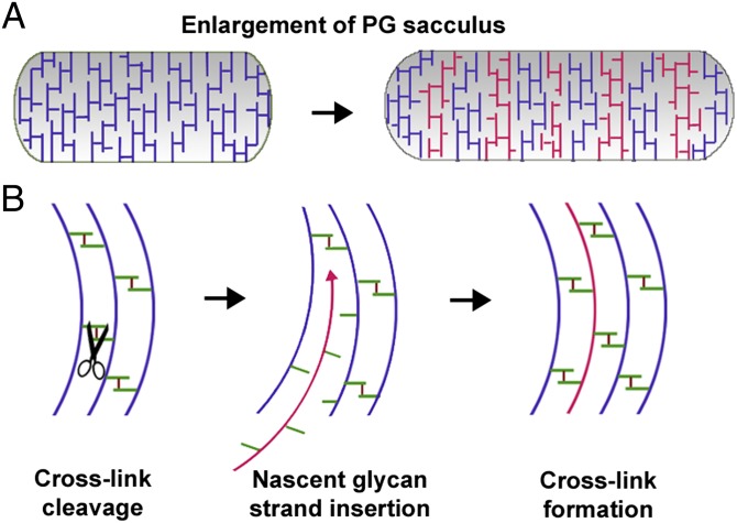
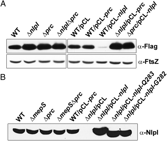
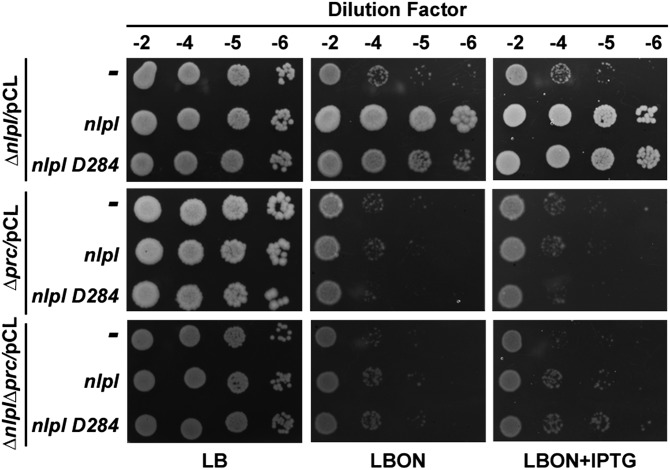
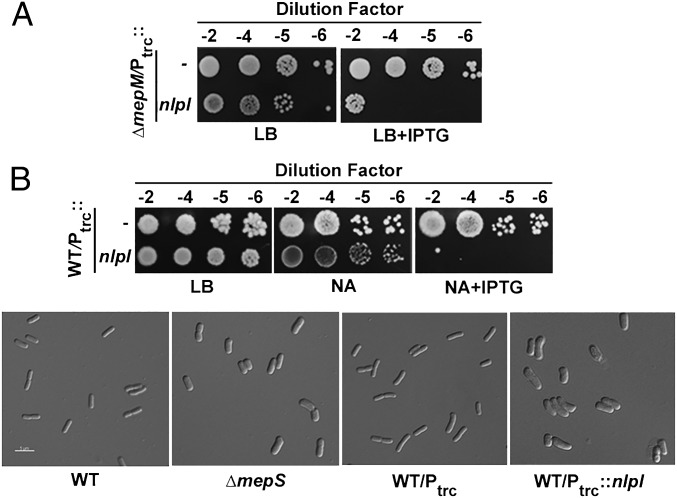
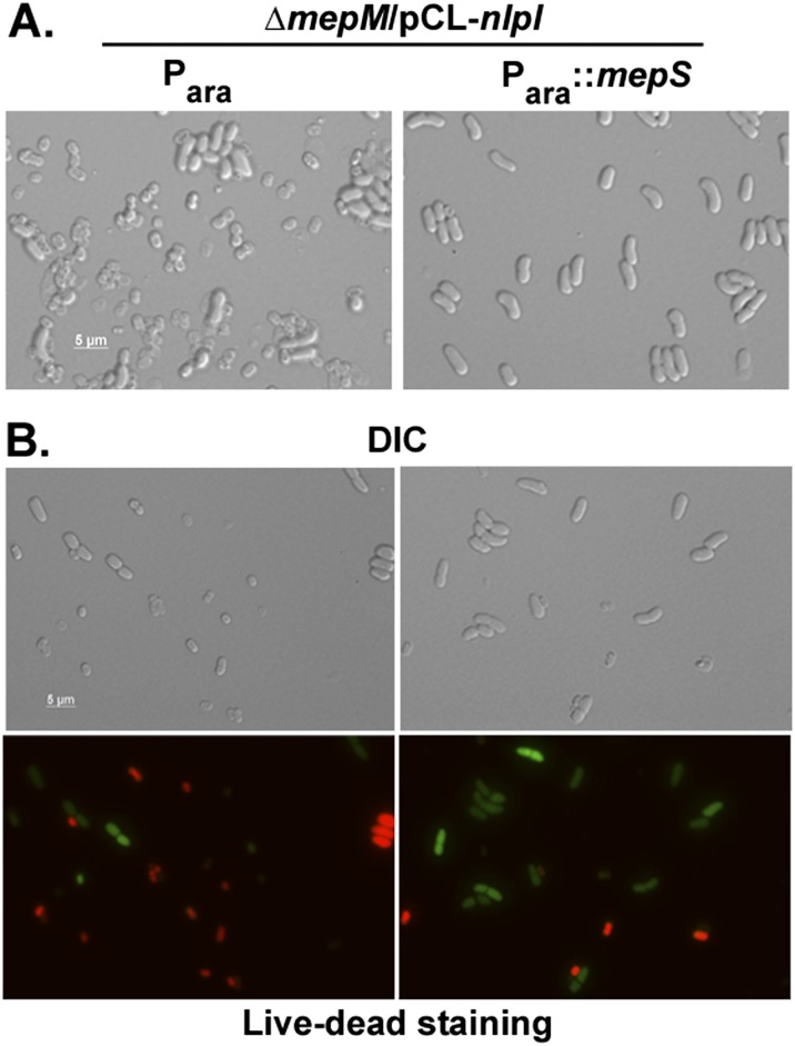
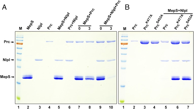
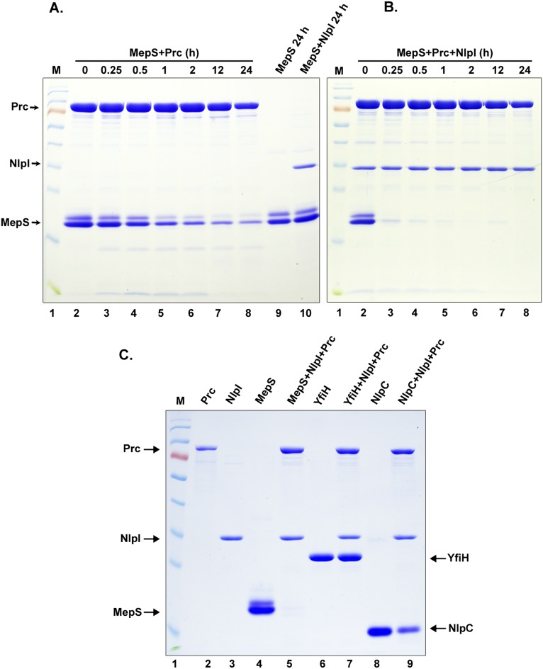
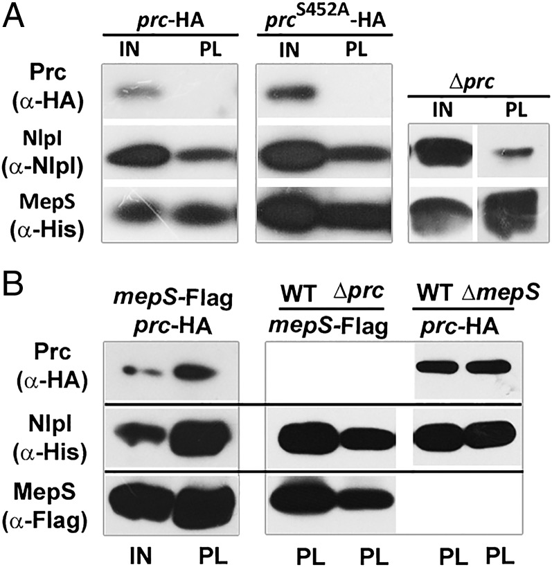
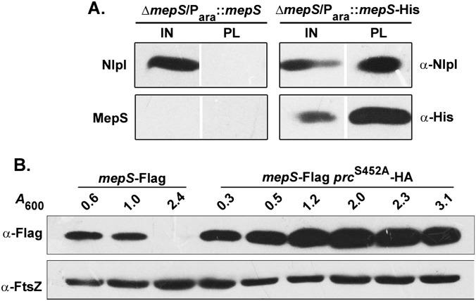
Bacterial growth and morphogenesis are intimately coupled to expansion of peptidoglycan (PG), an extensively cross-linked macromolecule that forms a protective mesh-like sacculus around the cytoplasmic membrane. Growth of the PG sacculus is a dynamic event requiring the concerted action of hydrolases that cleave the cross-links for insertion of new material and synthases that catalyze cross-link formation; however, the factors that regulate PG expansion during bacterial growth are poorly understood. Here, we show that the PG hydrolase MepS (formerly Spr), which is specific to cleavage of cross-links during PG expansion in Escherichia coli, is modulated by proteolysis. Using combined genetic, molecular, and biochemical approaches, we demonstrate that MepS is rapidly degraded by a proteolytic system comprising an outer membrane lipoprotein of unknown function, NlpI, and a periplasmic protease, Prc (or Tsp). In summary, our results indicate that the NlpI-Prc system contributes to growth and enlargement of the PG sacculus by modulating the cellular levels of the cross-link-cleaving hydrolase MepS. Overall, this study signifies the importance of PG cross-link cleavage and its regulation in bacterial cell wall biogenesis.

Keywords: MepS; NlpI-Prc; bacterial morphogenesis; peptidoglycan; regulated proteolysis.


Conflict of interest statement

The authors declare no conflict of interest.

Figures

Fig. 1.
Fig. 1.
Schematic of PG sacculus expansion. (A) Depiction of PG sacculus enlargement during growth of a bacterial cell. New murein strands (colored red) are incorporated into the preexisting murein strands (colored blue) to expand the PG sacculus (2, 3). (B) A model showing cleavage and resynthesis of cross-links for successful insertion of new glycan strands for expansion of the PG sacculus (8). The green bars indicate peptide stems whereas the small dark red bars represent the cross-bridges between the peptide stems of adjacent glycan strands.
Fig. 2.
Fig. 2.
Growth phase specificity of MepS. Strain MR802 (MG1655 mepS-Flag) was grown in LB and fractions were collected at various time intervals (with indicated A600 values) during the growth cycle. Normalized cell extracts (corresponding to 0.25 OD cells) were separated by SDS/PAGE, and MepS was detected by Western analysis using anti-Flag antisera. FtsZ was used as a loading control.
Fig. S1.
Fig. S1.
Regulation of mepS by NlpI and Prc. (A) Qualitative patch assay for activity of MepS-PhoA. Cells of MR807 (MG1655 ΔphoA ΔmepS), MR808 (MR807 ΔnlpI::Kan), or MR809 (MR807 Δprc::Kan) carrying plasmid pMN223 (Para::mepS-phoA) were patched onto minimal-121 plates containing 0.01% arabinose, 10−4 M KH2PO4, and 40 μg/mL 5-bromo-4-chloro-3-indolyl phosphate (BCIP) and grown at 37 °C. nlpI and prc mutants showed a marginal but consistent increase of the blue coloration, which is an indicator of MepS-PhoA activity. (B) The absence of mepS suppresses the growth defects of nlpI and prc mutants. Cells of WT (MG1655) and its various mutant derivatives were grown in LB broth, and 5 μL of the indicated dilutions were spotted on LB and LBON (LB without added NaCl) plates and grown at 42 °C. The genetic interactions between mepS, nlpI, and prc were described earlier (16, 17). (C) d,d-endopeptidase activity of MepS is lethal in nlpI and prc mutants. Cells of MR813 (ΔmepS ΔnlpI) or MR814 (ΔmepS Δprc) carrying pBAD33 (Para), pMN83 (Para::mepS), or pMN88 (Para::mepS-C68A) were grown in LB plus Cam broth, and 5 μL of various dilutions were spotted on indicated plates and grown at 42 °C.
Fig. 3.
Fig. 3.
Regulation of MepS is dependent on NlpI and Prc. (A) MepS-Flag levels in nlpI and prc mutants. Strains MR802 (MG1655 mepS-Flag), MR803 (MR802 ΔnlpI), or MR804 (MR802 Δprc) were grown in LB, and fractions were collected at different time points during growth and analyzed by Western blotting as described in the legend to Fig. 2. (B) Determination of stability of MepS by pulse–chase experiment (in vivo degradation assay). To rapidly growing cultures of the above strains in LB (at an OD600 of ∼0.4), spectinomycin (Spec) was added at a concentration of 300 μg/mL to block translation, and fractions were collected at indicated time points and analyzed as described in the legend to Fig. 2.
Fig. 4.
Fig. 4.
Requirement of both NlpI and Prc for regulation of MepS. (A) Levels of MepS in single and double mutants of nlpI and prc. Strains MR802 (MG1655 mepS-Flag), MR803 (MR802 ΔnlpI), MR804 (MR802 Δprc), and MR806 (MR802 ΔnlpI Δprc) were grown in LB to an A600 of 1.0, and MepS was detected by Western analysis as described in the legend to Fig. 2. Plasmid-carrying strains were grown with appropriate antibiotic (Spec) and 1 mM IPTG. FtsZ was used as a loading control. (B) NlpI is not processed by Prc. The indicated strains MG1655 (WT), MR810 (MG1655 ΔmepS), MR812 (MG1655 Δprc), MR814 (MG1655 ΔmepS Δprc) were grown in LB and harvested at an A600 of ∼1.0. Normalized cell extracts were analyzed by Western blotting using anti-NlpI antisera. Plasmid-carrying strains were grown with Spec and 1 mM IPTG. The C-terminal truncations of NlpI (bands in lanes 8 and 9) served as controls to indicate the size of processed NlpI. Processed NlpI is expected to be of 284 amino acids in length (17) whereas full-length is 294 amino acids.
Fig. S2.
Fig. S2.
Complementation using full-length nlpI or nlpI-D284 plasmid derivatives. Cells of MR811 (ΔnlpI), MR812 (Δprc), or MR815 (ΔnlpI Δprc) carrying pCL1920, pMN216 (pCL-nlpI), or pMN211 (pCL-nlpI-D284) were grown in LB broth with Spec, and 5 μL of various dilutions were spotted on indicated plates and grown at 42 °C. IPTG was used at 1 mM.
Fig. 5.
Fig. 5.
NlpI overexpression phenocopies MepS phenotype. (A) Lethal effect of overexpressed NlpI in ΔmepM mutants. Strains MR816 (BW27783 ΔmepM) carrying either pTRC99a (Ptrc) or pMN204 (Ptrc::nlpI) were grown in LB with Amp, and 5 μL of various dilutions were placed on indicated plates and grown. IPTG was used at 100 μM. (B) Effect of overexpressed NlpI in WT E. coli. MG1655 carrying either pTRC99a (Ptrc) or pMN204 (Ptrc::nlpI) was cultured and grown on nutrient agar (NA) at 42 °C. IPTG was used at 100 μM. ΔmepS strains are known not to grow on NA at 42 °C (24). For microscopy, the cultures were grown, diluted 1:100 either in LBON (for WT and ΔmepS strains) or in LBON plus 100 µM IPTG (for plasmid-carrying strains) and grown at 42 °C until an OD600 of ∼0.4–0.6. Differential interference contrast (DIC) images were taken after concentrating and spotting the culture on agarose pads. Overexpression of NlpI in WT is known to result in loss of rod shape and formation of prolate ellipsoid cells (15). It is not clear why the mutants lacking MepS (or those overproducing NlpI) exhibit fat and wide cell morphology; these cells may possibly be partially deficient in cross-link cleavage, leading to defective growth and elongation of the PG sacculus.
Fig. S3.
Fig. S3.
Effect of multiple copies of nlpI is suppressible by additional copies of mepS. (A) Strain MR816 /pMN216 carrying either pBAD33 (Para) or pMN83 (Para::mepS) was grown in LB with appropriate antibiotics and subcultured 1:100 in LB broth containing 0.2% arabinose, and, after 1 h growth (∼0.15 OD), 500 μM IPTG was added and grown for a further 1 h, after which cultures were drawn for microscopy. (B) Strains were grown as described above, and live–dead cell staining was done as described in SI Materials and Methods. Green and red represent live and dead cells, respectively. (Scale bars are shown.)
Fig. 6.
Fig. 6.
MepS is a substrate of Prc. (A) In vitro degradation assays. The C-terminal hexahistidine-tagged MepS, NlpI, and Prc proteins were mixed in all combinations (as indicated) and incubated for 3 h at 37 °C followed by SDS/PAGE and Coomassie blue staining. Each reaction contained the following (approximately): MepS, 15 μg; NlpI, 2 μg; and Prc, 4 μg. The mixtures in lanes 7 and 9 served as controls (0 h; no incubation). M is a protein marker with the following molecular size range (in kDa): 170, 130, 100, 70, 55, 40, 35, 25, 15, and 10. Purified MepS was always seen on SDS/PAGE as a doublet, and N-terminal sequencing confirmed that both bands belong to MepS itself. (B) Specificity of Prc. Two mutant derivatives of Prc (carrying K477A or S452A alterations at the active site residues) did not show any proteolytic activity (even when higher concentrations were used) against MepS in the presence of NlpI.
Fig. S4.
Fig. S4.
MepS is a substrate of Prc. (A) Time course degradation assay of MepS. Purified proteins (MepS and Prc) were mixed and incubated at 37 °C as indicated, followed by SDS/PAGE and Coomassie staining. MepS undergoes significant degradation on prolonged incubation with Prc alone (compare lanes 2 and 8), but not when incubated with NlpI (lanes 9 and 10). (B) Time course degradation assay of MepS. Purified proteins (MepS, NlpI, and Prc) were mixed and incubated as indicated at 37 °C, followed by SDS/PAGE and Coomassie staining. MepS degradation is enhanced quite considerably in the presence of NlpI (lanes 2 and 3). (C) Purified YfiH (a putative peptidase) or NlpC (a paralog of MepS) was used as a substrate of Prc. The proteins as indicated were added and incubated for 3 h at 37 °C, followed by SDS/PAGE and Coomassie staining. Lanes 6 and 8 show purified C-terminal His-tagged YfiH and NlpC proteins. YfiH is not degraded at all whereas NlpC seems to be degraded to some extent (lanes 7 and 9).
Fig. 7.
Fig. 7.
Interactions of MepS, NlpI, and Prc. (A) Western blot showing interaction of MepS-His with NlpI and Prc. Strains MR818 (MG1655 ΔmepS prc-HA-Cam), MR819 (MG1655 ΔmepS prcS452A-HA-Cam), or MR814 (MG1655 ΔmepS Δprc) carrying pMN217 (Para::mepS-His) were grown overnight in LB with Amp and next morning diluted 1:100 into fresh LB and at A600 of ∼0.2–0.3 expression of MepS was induced by addition of 0.05% l-arabinose. Cultures were further grown until an A600 of ∼0.8–1.0, and MepS-His was purified using Ni2+-NTA beads as described in SI Materials and Methods. The purified fractions of MepS-His (pull-down fractions, PL) along with the input samples (IN) were separated on SDS/PAGE, and immunoblot analysis was performed to detect MepS, NlpI, and Prc with anti-His, anti-NlpI, and anti-HA antibodies, respectively. All strains had a deletion of mepS to reduce the interference from the endogenous MepS protein. A strain carrying plasmid-borne, untagged MepS (MG1655 ΔmepS/Para::mepS) was used as a negative control (Fig. S5A). (B) Western blot showing interaction of NlpI-His with MepS and Prc. Strains MR805 (MG1655 ΔnlpI mepS-Flag), MR806 (MG1655 ΔnlpI Δprc mepS-Flag), MR820 (MG1655 ΔnlpI mepS-Flag prc-HA-Cam), MR821 (MG1655 ΔnlpI ΔmepS prc-HA-Cam), and MR822 (MG1655 ΔnlpI prc-HA-Cam) carrying pMN218 (Para::nlpI-His) were grown with 0.05% l-arabinose to an OD of 0.8–1.0. NlpI-His was purified from all these strains using an Ni2+-NTA column, and the purified fractions (PL) along with the input fractions (IN) were separated on SDS/PAGE. Western analysis was done using anti-His, anti-Flag, and anti-HA antibodies to detect NlpI, MepS, and Prc, respectively. In both these experiments, input sample corresponds to ∼0.3 OD cells whereas the pull-down fraction is ∼20-fold concentrated (i.e., from ∼6 OD cells).
Fig. S5.
Fig. S5.
Interactions of MepS with NlpI and Prc. (A) Western blot showing interaction of MepS with NlpI. Strains MG1655 ΔmepS carrying either Para::mepS or Para::mepS-His were grown with 0.05% arabinose as described in the legend to Fig. 7, and MepS was purified from both these strains using Ni2+-NTA beads. The purified fractions (PL) along with the input samples (IN) were separated by SDS/PAGE, and immunoblot analysis was performed to detect MepS and NlpI using anti-His or anti-NlpI antibodies. The untagged MepS did not show any interaction with NlpI, showing that the bands (NlpI) are specific for the MepS-His tag. (B) Western blot depicting the levels of MepS in WT and in prcS452A mutant. Strain MR802 and its prcS452A-HA-Cam derivative were grown in LB, fractions were collected at various time points, and normalized volumes of cell extracts were separated by SDS/PAGE followed by Western analysis using anti-Flag antibodies. FtsZ was used as loading control.
Fig. S6.
Fig. S6.
Localization of Prc. The subcellular localization of Prc was examined after partial cell fractionation using the MG1655 prc-Flag-Kan strain. Cells were incubated in Tris-sucrose plus lysozyme plus EDTA buffer, followed by brief sonication for cell lysis. Unbroken cells were removed by a low-speed centrifugation step, and the supernatant (total cell lysate) was centrifuged at 4 °C for 1 h at 1,00,000 × g. Resuspended pellet (membrane fraction) and the supernatant (soluble fraction) were loaded on SDS/PAGE along with the total cell lysate, and Western analysis was done to detect Prc using an anti-Flag antibody. The other marker proteins included Pbp1b and FtsI, which are IM-anchored periplasmic proteins, and MBP, a soluble periplasmic protein. The data indicate that Prc is a soluble protein.
Fig. S7.
Fig. S7.
Levels of NlpI and Prc during the bacterial growth cycle. The protein levels of NlpI and Prc were examined using MG1655 carrying a functional prc-Flag-Kan allele. This strain was grown in LB, fractions were taken at the indicated optical density, and normalized cell extracts were analyzed by Western blotting using anti-Flag, anti-NlpI, and anti-FtsZ antisera to detect Prc, NlpI, and FtsZ, respectively. FtsZ was used as a loading control. Strain MR802 (MG1655 mepS-Flag) was also used as a control.

References

    1. Weidel W, Pelzer H. Bagshaped macromolecules: A new outlook on bacterial cell walls. Adv Enzymol Relat Areas Mol Biol. 1964;26:193–232. - PubMed
    1. Park JT. The murein sacculus. In: Neidhardt FC, et al., editors. Escherichia coli and Salmonella: Cellular and Molecular Biology. 2nd Ed. ASM; Washington, DC: 1996. pp. 48–57.
    1. Höltje JV. Growth of the stress-bearing and shape-maintaining murein sacculus of Escherichia coli. Microbiol Mol Biol Rev. 1998;62(1):181–203. - PMC - PubMed
    1. den Blaauwen T, de Pedro MA, Nguyen-Distèche M, Ayala JA. Morphogenesis of rod-shaped sacculi. FEMS Microbiol Rev. 2008;32(2):321–344. - PubMed
    1. Typas A, Banzhaf M, Gross CA, Vollmer W. From the regulation of peptidoglycan synthesis to bacterial growth and morphology. Nat Rev Microbiol. 2012;10(2):123–136. - PMC - PubMed

Publication types

MeSH terms