. 2015 Sep 1;112(35):E4854-63.
doi: 10.1073/pnas.1509716112. Epub 2015 Aug 17.

Screen for multi-SUMO-binding proteins reveals a multi-SIM-binding mechanism for recruitment of the transcriptional regulator ZMYM2 to chromatin

Affiliations

Screen for multi-SUMO-binding proteins reveals a multi-SIM-binding mechanism for recruitment of the transcriptional regulator ZMYM2 to chromatin

Elisa Aguilar-Martinez et al. Proc Natl Acad Sci U S A. .

Abstract

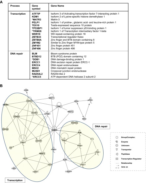
Protein SUMOylation has emerged as an important regulatory event, particularly in nuclear processes such as transcriptional control and DNA repair. In this context, small ubiquitin-like modifier (SUMO) often provides a binding platform for the recruitment of proteins via their SUMO-interacting motifs (SIMs). Recent discoveries point to an important role for multivalent SUMO binding through multiple SIMs in the binding partner as exemplified by poly-SUMOylation acting as a binding platform for ubiquitin E3 ligases such as ring finger protein 4. Here, we have investigated whether other types of protein are recruited through multivalent SUMO interactions. We have identified dozens of proteins that bind to multi-SUMO platforms, thereby uncovering a complex potential regulatory network. Multi-SUMO binding is mediated through multi-SIM modules, and the functional importance of these interactions is demonstrated for the transcriptional corepressor ZMYM2/ZNF198 where its multi-SUMO-binding activity is required for its recruitment to chromatin.

Keywords: SIM; SUMO; ZMYM2; ZNF198; chromatin.


Conflict of interest statement

The authors declare no conflict of interest.

Figures

Fig. 1.
Fig. 1.
Identification of multi-SUMO–binding proteins. (A) Schematic representation of potential mechanisms through which a multi-SIM–containing protein can interact with a SUMOylated substrate (X) through binding to poly-SUMO chains (Left) or a multiple SUMO molecules (S) present on a platform of several mono-SUMOylated lysine (K) residues (Right). (B) Schematic representation of GST-SUMO and “pentameric” GST-COMP-SUMO. (C) The indicated purified GST-fused proteins were resolved by denaturing (Top) or native (Bottom) polyacrylamide gel electrophoresis. Proteins were visualized with Coomassie blue stain. (D and E) GST pulldown assays of WT or mutant MBP-RNF4 with GST or the indicated GST-fusion proteins. (D) RNF4 binding is shown at the Top using an anti-MBP antibody for immunoblot (IB), and a Ponceau-stained membrane at the Bottom shows the GST bait input proteins. The 5% input protein is shown (lane 1). (E) Coomassie-stained gel showing the GST-COMP-SUMO bait proteins and pulled down (PD) WT and SIM2 mutant versions of MBP-RNF4. (F) Strategy for identifying multi-SUMO interacting proteins, from nuclear extracts by binding to GST-COMP-SUMO3. (G) Coomassie-stained gel showing bait proteins (lanes 3 and 4) and proteins pulled down by GST-COMP and GST-COMP-SUMO3. Molecular-weight markers (M1 and M2) are indicated. (H) Pie chart showing the cellular function of the GST-COMP-SUMO3 binding proteins found by mass spectrometry.
Fig. S1.
Fig. S1.
Paralog specificity of RNF4 binding to multi-SUMO scaffolds. GST-pulldown assay of FLAG-tagged RNF4 (expressed in HEK293T cell lysates) with the indicated GST-tagged COMP fusion proteins. Proteins pulled down (PD) (lanes 5–7) or the unbound protein in the flow through (FT) (lanes 2–4) were detected by IB with an anti-FLAG antibody (Top). Proteins used as baits are shown by Coomassie stain at the Bottom. The 5% input protein is shown (lane 1). RNF4 shows specificity toward binding to SUMO3 in the context of the GST-COMP-SUMO3 multi-SUMO scaffold.
Fig. S2.
Fig. S2.
Interaction network of proteins found to interact with multi-SUMO by mass spectrometry. (A) Proteins involved in transcription or DNA repair that were identified by mass spectrometry after binding to the GST-COMP-SUMO3 multi-SUMO scaffold. Proteins denoted with an asterisk were from the lower confidence dataset. (B) A network of known interactions was constructed using Ingenuity Systems software based on using SUMO as the starting protein and all of the proteins identified as binding to the multi-SUMO scaffold. Parts of the network associated with transcriptional control and DNA repair are highlighted.
Fig. 2.
Fig. 2.
Validation of multi-SUMO–binding interactions. (A) GST pulldown analysis of PTRF, BLM, and ZMYM2 binding to the indicated GST-fusion proteins. Epitope-tagged PTRF, BLM, or ZMYM2 were overexpressed in HEK293T cells, and total cell lysates were used for the pulldown. Proteins were detected by IB using anti-FLAG (PTRF) or anti-Myc (BLM and ZMYM2) antibodies. Arrows indicate the positions of bands corresponding to the full-length proteins. Bait proteins were stained with Ponceau (Bottom). (B) Interaction of ZBTB33 or ZBTB4 with COMP-SUMO3 was analyzed by GST-pulldown. Total cell extracts of transfected HEK293T cells were used for the pulldown. Input and precipitated ZBTB33 and ZBTB4 were detected using an anti-FLAG antibody. Proteins used as bait were stained with Ponceau (Bottom).
Fig. S3.
Fig. S3.
Multi-SUMO interactions with SRBC and ZMYM2. (A) The interaction of SRBC with the indicated GST-fusion proteins was analyzed by GST pulldown using total cell extracts of HEK293T cells transfected with a myc-tagged SRBC expression construct. SRBC was detected by IB using an anti-myc antibody. A long exposure of the blot is presented here, showing low-level nonspecific binding of SRBC to the GST-COMP fusion proteins. The input sample lane (5% input) shows saturation (lane 1; gray line). (B) GST pulldown analysis of the interaction of myc-tagged SRBC with the indicated GST-fusion proteins when coexpressed with PTRF in HEK293T cell lysates. The binding of PTRF when expressed alone to the different GST-fusion constructs is shown in Fig. 2A. Specific binding of SRBC to GST-COMP-SUMO3 is revealed in the presence of coexpressed PTRF. (C) In vitro-translated ZBTB33 was used in a GST pulldown assay with the indicated GST-fusion proteins. Bait proteins were stained with Coomassie (Bottom), and ZBTB33 was detected by autoradiography (Top). In all cases, 10% input protein is shown. (D, Left) Coomassie-stained SDS/PAGE of the purified GST and GST-ZMYM2 input proteins. Arrow indicates the position of full-length GST-ZMYM2(1–200). (Right) IB of recombinant purified His-tagged COMP (Top) or COMP-SUMO3 (Bottom) following pulldown with GST or GST-ZMYM2(1–200). Proteins were detected using an anti-His antibody. The 5% input is shown (lane 1). Direct specific interactions are observed between ZMYM2 and the multimeric SUMO3 scaffold. (E) GST pulldown analysis of the interaction of purified recombinant GST-ZMYM2(1–200) with multi or poly forms of His-tagged SUMO. Poly-SUMO is presented in the form of linear fusion proteins. (Top) Schematic illustration of the constructs used for the analysis. (Bottom Left) Coomassie-stained SDS/PAGE of the His-tagged purified proteins. (Bottom Right) Proteins binding to ZMYM2 are shown by immunoblot (IB) with anti-His antibody (Top), and the GST bait proteins are shown on a Ponceau-stained membrane (Bottom). (F) Binding of SUMO domain proteins to immobilized control protein GST-Kistrin by SPR. At t = 0, either COMP (gray sensorgram), COMP-SUMO3 (blue sensorgram), or SUMO3×4 (orange sensorgram) was injected. (G) Immunoprecipitation (IP) analysis of ZMYM2 from U2OS-Flp-In cells containing doxycycline (Dox)-inducible FLAG-tagged SUMO3(K11R/Q90P) in the absence and presence of Dox. Coprecipitated SUMO3 was detected by IB with anti-FLAG antibody.
Fig. 3.
Fig. 3.
Characterization of SIM–multi-SUMO binding interactions. (A) Structure of SUMO3 indicating the surface involved in the interaction with SIMs (41). Hydrophobic amino acids are shown in black; basic amino acids are colored blue. The locations of three residues in the surface that we mutated to alanine to disrupt SIM interactions are shown in yellow (Right). The sequences surrounding these amino acids in wild-type (WT) SUMO3 or the triple-alanine mutant SUMO3(3A) are shown below. Mutated amino acids are in bold. (B–E) GST pulldown (PD) assays with the indicated GST-fusion proteins. After the PD, bait proteins were stained with Ponceau (Bottom). (B) Binding of RNF4 or ZMYM2 (from lysates of HEK293T cells overexpressing the proteins) to WT GST-COMP-SUMO3 or GST-COMP-SUMO3(3A). Interacting proteins were detected by IB with anti-FLAG (RNF4) or anti-myc (ZMYM2) antibodies (Top). (C) Interaction of FLAG-tagged ZBTB33, PTRF, PIAS1, or SENP3cs from lysates of HEK293T cells overexpressing these proteins with WT SUMO3 or SUMO3(3A). Proteins were detected by immunoblot using anti-FLAG antibodies (Top). (D and E) Peptide competition assays. Bait proteins were stained with Ponceau (Bottom). (D, Top) Sequence of the SIM peptide used for the competition assays. Acidic amino acids are underlined, and the hydrophobic core boxed. (Bottom) IB using anti-FLAG antibody showing RNF4 binding to GST-COMP-SUMO3 after incubation with increasing concentrations of SIM peptide (0, 0.5, 5, and 50 ng). RNF4 was from cell lysates of transfected HEK293T cells. (E) Assay was carried out as described in D, but only 0, 25, and 50 ng of SIM peptide were used and the cell lysates were from HEK293T cells expressing catalytically inactive SENP3cs or ZMYM2. Interacting proteins were detected by IB using an anti-FLAG (for SENP3CS) or anti-myc (for ZMYM2) antibody.
Fig. 4.
Fig. 4.
Mapping the SIMs in ZMYM2. (A) Schematic illustration of ZMYM2 showing the location and sequence of three putative SIMs. Acidic amino acids are colored in red. Regions containing hydrophobic amino acids mutated to alanine are boxed. (B) GST pulldown analysis of the indicated GST-fusion proteins to WT or mutant forms of ZMYM2. Total cell lysates of HEK293T cells overexpressing the ZMYM2 proteins were used for the PD. ZMYM2 was detected by IB using anti-myc antibody. The 5% input is shown. Quantification of ZMYM2 binding to GST-COMP-SUMO3 is shown on the Right as a percentage relative to the input lane and is the average of two experiments. Error bars represent SE. (C) GST pulldown analysis of the interaction of the purified recombinant GST-fusion proteins, COMP, COMP-SUMO3 (multi-SUMO), and SUMO3×4 (poly-SUMO is presented in the form of a linear fusion protein) with the indicated proteins from transiently transfected HEK293T cell lysates. (Top) Schematic illustration of the constructs used for the analysis. (Bottom) Proteins binding to the indicated GST-fusion proteins are shown by immunoblot (IB) with antibodies to the specific tags (Fig. 2) (top panels) and the GST bait proteins are shown on a Ponceau-stained membrane (bottom panel). Quantification of binding of each protein to each of the GST-fusion proteins is shown on the Right as a percentage relative to binding to GST-SUMO3×4 (taken as 100%). Data are the average of three experiments, and error bars represent SD. (D) GST pulldown analysis of GST-COMP-SUMO3 or GST-SUMO3×4 to WT or mutant forms of ZMYM2. Total cell lysates of HEK293T cells overexpressing the ZMYM2 proteins were used for the PD. ZMYM2 was detected by IB using anti-myc antibody. The 5% input is shown. (E) Binding of the indicated SUMO fusion proteins to immobilized GST-ZMYM2(1–200) by SPR. At t = 0, either COMP (gray sensorgram), COMP-SUMO3 (blue sensorgram), or SUMO×4 (orange sensorgram) was injected. Dissociation rates (kd) were 6.05 ± 1.11 × 10⁻⁴ s−1 (measured between 200 and 300 s) for COMP-SUMO3 and 7.51 ± 1.64 × 10⁻² s−1 (measured between 62 and 100 s) for SUMO3×4. The kd values are mean ± SD from three separate experiments.
Fig. S4.
Fig. S4.
The SIMs of ZMYM2 are important for transcriptional regulation. (A) Immunofluorescence of transiently transfected U2OS cells expressing WT, SIM2mut, or SIM1,2,3mut myc-tagged ZMYM2. Proteins were detected using an anti-myc primary and anti-mouse Alexa Fluor 594 secondary antibodies, and DNA was detected by Hoechst staining. Projections of deconvolved images and a merge of the two signals (DNA, blue, and ZMYM2, red) are shown. (B) Expression of wild-type (WT) or the SIM2 mutant form of ZMYM2 was detected by IB with anti-FLAG antibodies. Stably transfected U2OS cells were treated with doxycycline (Dox) for 48 h, where indicated. Lamin B was detected by IB as loading control. (C) Immunoblot of ZMYM2 expression in the parental U2OS Flp-In cell line or U2OS cells stably transfected with constructs expressing WT, SIM2mut, or SIM1,2,3mut and treated for 48 h with doxycycline. Proteins were detected using anti-ZMYM2 (Top) or anti-FLAG (Bottom) antibodies. Gray arrows indicate the endogenous protein, black arrows indicate the exogenous FLAG-tagged ZMYM2, and the asterisk indicates an unspecific band. (D) RT-PCR analysis of the expression of the indicated genes in U2OS cells stably expressing the SIM2 mutant form of ZMYM2. Data are the average of two (for TMEM154) or three independent experiments and are presented as log10 of the fold change in ZMYM2(SIM2mut) cells relative to the transcript levels in WT ZMYM2-expressing cells (taken as 1). Asterisks (*) denote P < 0.05 in Student’s t test and a fold change >5. (E) RT-PCR analysis of the expression of the indicated genes in U2OS cells stably expressing the SIM1,2,3 mutant form of ZMYM2. Data are the average of three independent experiments and are presented as log10 of the fold change relative to the transcript levels in WT ZMYM2-expressing cells (taken as 1). Asterisks (*) denote P < 0.05 in Student’s t test and a fold change >1.6.
Fig. S5.
Fig. S5.
Effect of ZMYM2 and UBE2I depletion on ZMYM2 target gene expression. (A) IB analysis of ZMYM2 expression in U2OS Flp-In cells following depletion with siZMYM2 and a nontargeting (siNT) control. Lamin B was used as loading control. (B) RT-PCR analysis of the expression of the indicated genes in U2OS cells transfected with control siNT (dark bars) or a siRNA duplex directed against ZMYM2 (lighter bars). Data are the average of three independent experiments and are presented as fold change relative to the transcript levels in control siNT-transfected cells (taken as 1). Asterisks (*) denote P < 0.05 in Student’s t test and a fold change >1.5. (C) IB analysis of UBC9/UBE2I expression following depletion with siUBC9/UBE2I or a nontargeting (siNT) control. Tubulin was used as loading control. (D) RT-PCR analysis of the expression of the indicated genes in U2OS Flp-In cells transfected with control siNT (dark bars) or a siRNA duplex directed against UBC9/UBE2I (lighter bars). Data are the average of two (for TMEM154) or three independent experiments and are presented as fold change relative to the transcript levels in control siNT-transfected cells (taken as 1). Asterisks (*) denotes P < 0.05 in Student’s t test and a fold change >1.5.
Fig. 5.
Fig. 5.
ZMYM2 SIMs are required for its recruitment to chromatin. (A) Reporter gene assay with constant LexA-VP16 but increasing amounts of the indicated Gal4-ZMYM2 derivatives. A schematic of the reporter gene is shown above, and the expression of the different Gal4 fusion proteins is shown in the Western blot as an Inset. Lamin B is a loading control, and the vertical line indicates that an intervening irrelevant lane has been removed. Data are shown relative to the internal β-galactosidase control (normalized to Gal4 DBD alone, taken as 1) and are the mean of two independent experiments, each performed in triplicate. (B) University of California, Santa Cruz genome browser view of three example loci and the binding peaks of the indicated ZMYM2 derivatives. (C) ChIP analysis of ZMYM2(WT) or ZMYM2(SIM2mut) binding to the indicated loci in the respective stable U2OS cell lines. Data are presented relative to input for each locus (n = 3), and statistically significant differences are indicated (P value <0.005). Control ChIPs with IgG are shown at each locus for each cell line.
Fig. 6.
Fig. 6.
Multi-SUMO binding promotes ZMYM2 recruitment to chromatin. (A) Heat map of tag densities from ZMYM2, SUMO, or control IgG ChIP-seq experiments plotted around 5 kb either side of the ZMYM2 binding peak summits. Binding regions are ranked according to ZMYM2 tag density. (B) Average tag density profiles of SUMO binding plotted onto 1-kb regions surrounding the summits of the top 1,000 most enriched peaks for either ZMYM2 or FOXO3. (C) Enrichment of SUMO associated with WT ZMYM2 binding relative to ZMYM2 SIM2mut binding. Total SUMO tags are plotted for the top 10% most ZMYM2(WT) tag enriched and the bottom 10% binding regions relative to ZMYM2mut binding. (D) ChIP analysis of SUMO2/3 (Left) or ZMYM2 (Right) binding to the indicated loci in U2OS cells at either 37 °C (black bars) or 43 °C (gray bars). Data are presented relative to input for each locus (n = 3) and statistically significant differences are indicated (value of P < 0.005). (E) Model showing different possible modes of SUMO (S) interactions with the multi-SIM module in ZMYM2 on chromatin. ZMYM2 could interact with multi-SUMOylated repressor (Rep) or corepressor (CoRep) proteins, multiple SUMO motifs found in just one protein, or multiple SUMO motifs found in different SUMOylated proteins including core histones.

References

    1. Heun P. SUMOrganization of the nucleus. Curr Opin Cell Biol. 2007;19(3):350–355. - PubMed
    1. Finkbeiner E, Haindl M, Raman N, Muller S. SUMO routes ribosome maturation. Nucleus. 2011;2(6):527–532. - PubMed
    1. Barry J, Lock RB. Small ubiquitin-related modifier-1: Wrestling with protein regulation. Int J Biochem Cell Biol. 2011;43(1):37–40. - PubMed
    1. Cubeñas-Potts C, Matunis MJ. SUMO: A multifaceted modifier of chromatin structure and function. Dev Cell. 2013;24(1):1–12. - PMC - PubMed
    1. Garcia-Dominguez M, Reyes JC. SUMO association with repressor complexes, emerging routes for transcriptional control. Biochim Biophys Acta. 2009;1789(6-8):451–459. - PubMed

Publication types

MeSH terms

LinkOut - more resources