. 2015 Sep 1;112(35):11007-12.
doi: 10.1073/pnas.1514730112. Epub 2015 Aug 17.

RIP3-mediated necrotic cell death accelerates systematic inflammation and mortality

Affiliations

RIP3-mediated necrotic cell death accelerates systematic inflammation and mortality

Lingjun Meng et al. Proc Natl Acad Sci U S A. .

Erratum in

Abstract

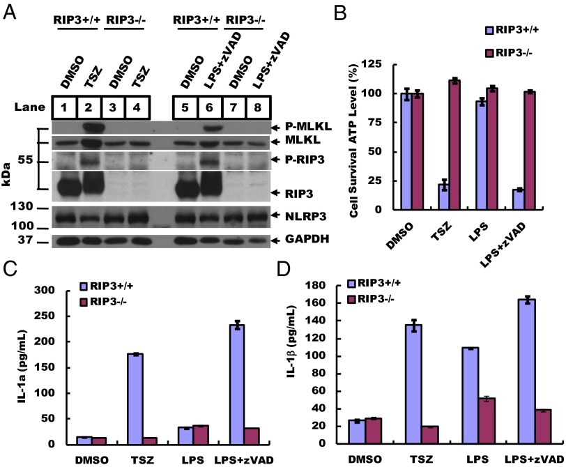
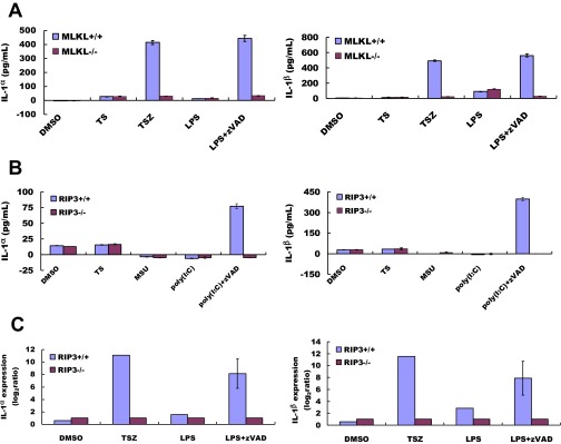
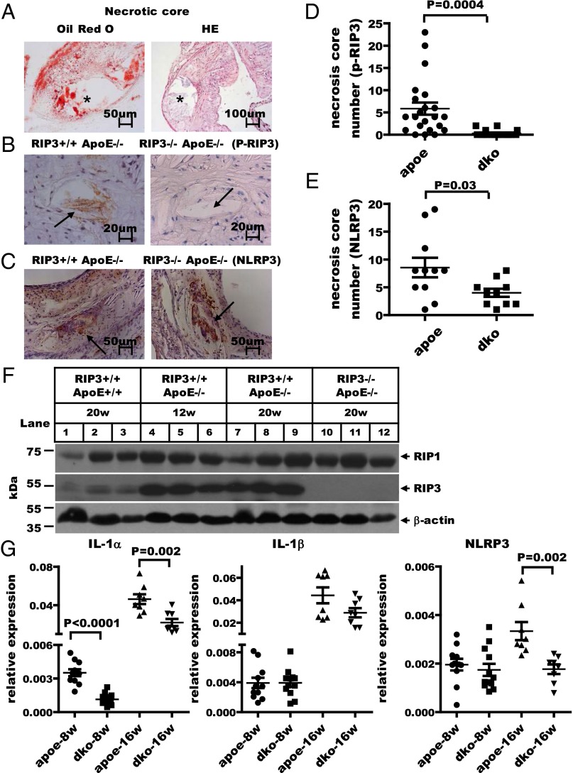
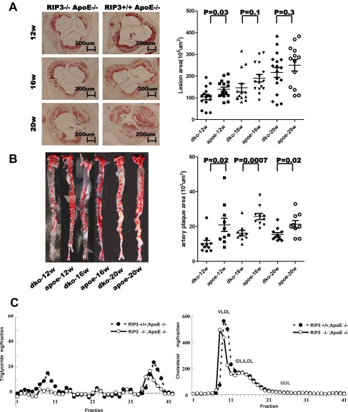
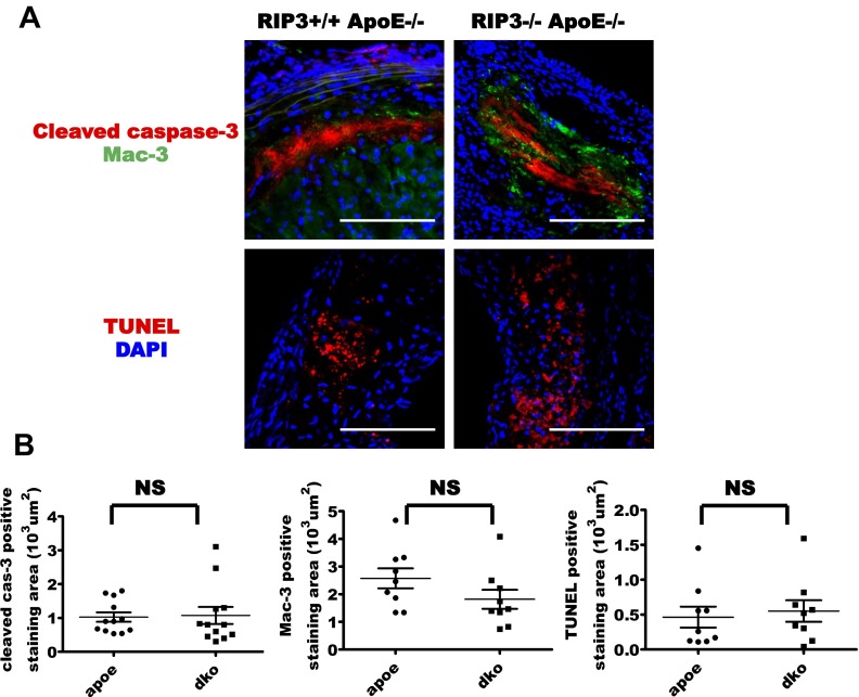
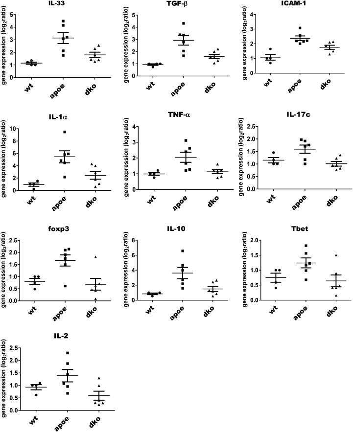
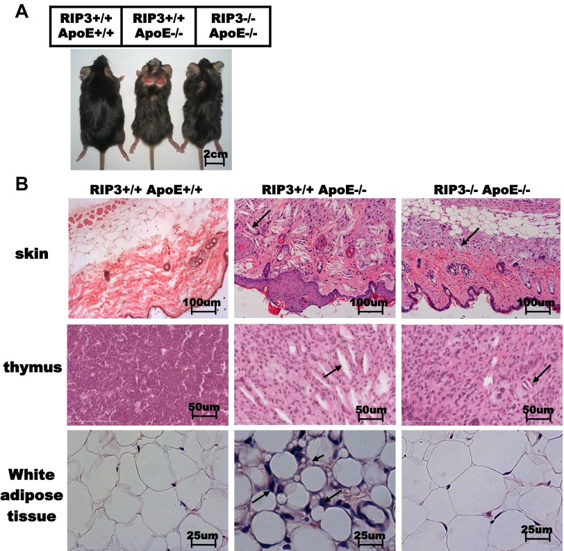
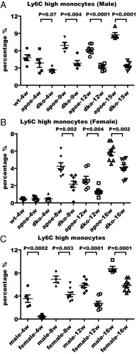
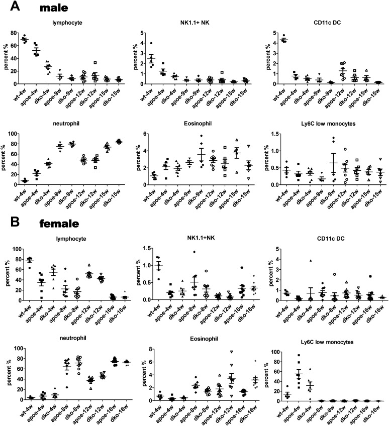
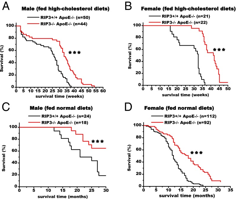
Systematic inflammation contributes to the development of many diseases, including cardiovascular disease, which is the leading cause of mortality worldwide. How such inflammation is initiated and maintained throughout the course of disease remains unclear. In the current study, we report the observation of specific phosphorylation of the receptor-interacting protein 3 (RIP3) kinase that marks the activation of programmed necrosis (also called the "necroptosis pathway") in the atherosclerotic plaques in apolipoprotein E (ApoE)-knockout mice. The mRNA expression levels of 10 inflammatory cytokines, including IL-1α, were decreased significantly in the plaque regions of mice lacking RIP3. Lymphocyte infiltrations in the adipocyte tissue and in skin lesions of ApoE single-knockout mice were significantly mitigated in ApoE/RIP3 double-knockout mice. The high percentage of inflammatory monocytes with high levels of lymphocyte antigen 6C in the blood of ApoE single-knockout mice also was greatly decreased in the ApoE/RIP3 double-knockout mice. Most significantly, the double-knockout mice displayed dramatically delayed mortality compared with ApoE single-knockout mice. Our findings indicate that necrotic death in areas such as atherosclerotic plaques may release cytokines that mobilize monocytes from bone marrow to the lesion sites, exacerbating the lesions in multiple tissues and resulting in the premature death of the animals.

Keywords: RIP3; atherosclerosis; longevity; macrophages; necrosis.


Conflict of interest statement

The authors declare no conflict of interest.

Figures

Fig. 1.
Fig. 1.
RIP3 deletion suppresses necrosis and IL-1α/β production. (A) BMDMs from 8-wk-old RIP3+/+ and RIP3−/− mice were treated with TSZ (20 ng/mL TNF-α, 100 nM Smac mimetic, and 20 µM zVAD) or LZ (20 ng/mL LPS and 20 µM zVAD) for 5 h. DMSO was used as a control. Cell lysates were collected, and sample aliquots were subjected to Western blot analysis of necrosis protein levels at the indicated conditions. Thirty micrograms of cell supernatant was loaded in each lane, except that 60 µg was loaded for phosphorylated RIP-3 (p-RIP3) and phosphorylated MLKL (p-MLKL). GAPDH is shown as a loading control. Blots were exposed to film at room temperature for 60 s. (B) BMDMs were treated at the indicated conditions for 12 h, and cell viability was determined by measuring ATP levels using a CellTiter Glo assay. Data represent the mean ± SD of duplicate samples. (C and D) BMDMs were cultured with different stimuli, as indicated, for 12 h. Secreted IL-1α and IL-1β levels were quantified by ELISA. All experiments were repeated at least three times with similar results.
Fig. S1.
Fig. S1.
RIP3 deletion suppresses necrosis and cytokine production at the mRNA and protein level. (A) Wild-type and MLKL−/− BMDMs were cultured in the indicated conditions (TS: 20 ng/mL TNF-α, 100 nM Smac mimetic; TSZ: 20 ng/mL TNF-α, 100 nM Smac mimetic, 20 mM zVAD; LPS: 20 ng/mL). Secreted IL-1α and IL-1β were quantified by ELISA. (B) BMDMs were cultured with TS for 12 h to induce apoptosis. DMSO was used as the untreated control. We treated BMDMs with the inflammation inducer MSU (300 μg/mL) and poly (I:C) (1 μg/mL) and added zVAD to induce necrosis. Secreted IL-1α and IL-1β were quantified by ELISA. Values are the mean ± SD of at least three experiments from different donors. (C) Real-time PCR analysis of IL-1α and IL-1β mRNA transcript levels in BMDMs stimulated with the indicated conditions as in A. Values presented are relative to the baseline expression in RIP3−/−, set as 1. Expression levels were normalized to GAPDH. Data represent the mean ± SD.
Fig. 2.
Fig. 2.
RIP3 deletion suppresses inflammation in atherosclerotic plaques. (A) Cross-sections of typical atherosclerotic lesions exhibiting extensive staining with Oil Red O and H&E. The asterisk indicates the necrotic core. (B) Representative IHC staining of p-RIP3 in plaques from ApoE single-knockout and RIP3/ApoE double-knockout mice fed high-cholesterol diets (1.25%) for 16 wk. Positive-staining signals appear as brown dots in sections counterstained with hematoxylin (blue). Plaque staining of phosphorylated RIP3 is seen only in the necrotic core of ApoE single-knockout mice (black arrow). (C) IHC staining representative of NLRP3 expression in necrotic cores from ApoE single-knockout and RIP3/ApoE double-knockout mice fed high-cholesterol diets for 16 wk. The black arrows indicate necrotic cores with NLRP3 staining. (D and E) Quantitative data showing the number of necrotic cores stained by phosphorylated RIP3 (n = 22 ApoE single-knockout mice; n = 20 RIP3/ApoE double-knockout mice) and NLRP3 (n = 11 ApoE single-knockout mice; n = 10 RIP3/ApoE double-knockout mice). Vertical bars show the mean ± SD for each group. dko, double knockout. (F) Western blot analysis of RIP1 and RIP3 levels in plaque areas microdissected from aortic valves/aortas of wild-type and RIP3/ApoE double-knockout mice fed high-cholesterol diets for 20 wk (n = 3) and of ApoE single-knockout mice fed the same diet for 12 wk. β-Actin is shown as a loading control. An 60-µg aliquot of atherosclerotic plaque extracts was loaded in each lane. All experiments were repeated at least three times with similar results. (G) Microdissected atherosclerotic plaque from aortic valves/aortas of ApoE single-knockout and RIP3/ApoE double-knockout mice fed high-cholesterol diets for 8 or 16 wk; mRNA was extracted for quantitative RT-PCR analysis of the expression of the inflammatory genes IL-1α, IL-1β, and NLRP3. The gene-expression levels were normalized to the expression level of GAPDH. Data represent the mean ± SD. Unpaired Student's t tests were used to evaluate significance. A P value < 0.01 was considered significant.
Fig. S3.
Fig. S3.
RIP3 deletion attenuated atherosclerotic lesion formation and had no significant effect on the levels of plasma cholesterol or triglycerides. (A, Left) Representative photographs of the aortic valve from RIP3/ApoE double-knockout and RIP3+/+ApoE−/− mice. Sections were stained with Oil Red O. (Right) Quantification of the atherosclerotic plaque area in cross-sections of proximal aorta in mice. Each symbol represents the lesion size in serial cross-sections from an individual male mouse fed a high-cholesterol diet for 12, 16, or 20 wk. (B, Left) Lesion size in the aorta. Representative Oil Red O staining of the inner plaque of aorta sections. (Right) Quantification of the atherosclerotic lesion area in en face analysis of the total aorta in mice. (C) Pooled plasma samples were subjected to FPLC gel filtration, and the fractions were assayed for cholesterol and triglyceride.
Fig. S2.
Fig. S2.
RIP3 deletion did not affect apoptosis in atherosclerotic plaques. (A) Sections from plaques from ApoE single-knockout and RIP3/ApoE double-knockout mice fed a high-cholesterol diet for 16 wk were stained with antibodies specific for cleaved caspase-3, macrophages (Mac-3), and TUNEL. Results are representative of immunofluorescence in 12 mice of each group. (Scale bars, 25 µm.) (B) Quantitative data showing the area stained by cleaved caspase-3, Mac-3, and TUNEL. Vertical bars show the mean ± SD per group. NS, not significant.
Fig. S4.
Fig. S4.
RIP3 deficiency decreased mRNA expression levels of cytokines in atherosclerotic ApoE background mice. Quantitative RT-PCR analysis of the relative gene mRNA expression in plaque microdissected from the aortic valves and aortas of mice fed a high-cholesterol diet for 16 wk (P < 0.05). The gene-expression levels were normalized to the expression of GAPDH. Data represent the mean ± SD. Unpaired Student's t tests were used to calculate significance. P < 0.05 was considered significant.
Fig. S5.
Fig. S5.
RIP3 deletion mitigated the inflammatory process. (A) Photographs of wild-type, ApoE single-knockout, and RIP3/ApoE double-knockout mice fed a high-cholesterol diet (1.25%) for 16 wk. (B) H&E-stained histological sections from skin and thymus (arrows indicate cholesterol crystals) and epididymal white adipose tissue (long arrows indicate the infiltrating cells that are most likely macrophages; short arrows indicate the small size of the adipocytes) of wild-type, ApoE single-knockout, and RIP3/ApoE double-knockout mice fed a high-cholesterol diet for 16 wk.
Fig. 3.
Fig. 3.
Deletion of RIP3 decreases the percentage of Ly6Chi inflammatory monocytes in blood during the progression of atherosclerotic disease. (A and B) Flow cytometry analysis of the percentage of the inflammatory monocyte population in the blood of male (A) and female (B) mice at the indicated times after high-cholesterol diet feeding. wt, wild type; apoe, ApoE single-knockout mice; dko, ApoE/RIP3 double-knockout mice. Cells were stained with antibodies against CD4, CD8, CD19, NK1.1, CD11c, CD11b, Ly6C, and Ly6G and were processed as described in Materials and Methods. (C) Comparison of the percentage of Ly6Chi inflammatory monocytes in male and female ApoE single-knockout mice.
Fig. S6.
Fig. S6.
RIP3 deletion affected Ly6Chi inflammatory monocytes but had no effect on other types of immune cells. (A and B) Flow cytometry analysis of the percentage of inflammatory monocyte populations in the blood of male (A) and female (B) mice during the progression of atherosclerotic disease. Cells were stained with antibodies against CD4, CD8, CD19, NK1.1, CD11c, CD11b, Ly6C, and Ly6G.
Fig. 4.
Fig. 4.
Deletion of RIP3 prolongs the lifespan of the atherosclerotic mice. (A and B) Comparison of survival of ApoE single-knockout and RIP3/ApoE double-knockout male (A) and female (B) mice. Survival curves start with 8-wk-old mice fed high-cholesterol diets (1.25%). ApoE single-knockout mice exhibited significantly decreased survival relative to RIP3/ApoE double-knockout mice. (C and D) Survival curves of ApoE single-knockout and RIP3/ApoE double-knockout male (C) and female (D) mice fed normal diets and recorded age at death. Data were analyzed with Kaplan–Meier tests. Asterisks denote the level of statistical significance between ApoE single-knockout and RIP3/ApoE double-knockout male and female mice: ***P < 0.001.
Fig. S7.
Fig. S7.
RIP3 deletion attenuated Ly6Chi inflammation monocytes and had no significant effect on the levels of estrogen. Serum estradiol and progesterone levels in RIP3+/+ApoE−/− and RIP3/ApoE double-knockout female mice fed high-cholesterol diets for 16 wk were evaluated by RIA.

References

    1. Libby P. Inflammation in atherosclerosis. Nature. 2002;420(6917):868–874. - PubMed
    1. Breslow JL. Mouse models of atherosclerosis. Science. 1996;272(5262):685–688. - PubMed
    1. Cohen I, et al. Differential release of chromatin-bound IL-1alpha discriminates between necrotic and apoptotic cell death by the ability to induce sterile inflammation. Proc Natl Acad Sci USA. 2010;107(6):2574–2579. - PMC - PubMed
    1. Thorp E, et al. Reduced apoptosis and plaque necrosis in advanced atherosclerotic lesions of Apoe-/- and Ldlr-/- mice lacking CHOP. Cell Metab. 2009;9(5):474–481. - PMC - PubMed
    1. Kamari Y, et al. Reduced atherosclerosis and inflammatory cytokines in apolipoprotein-E-deficient mice lacking bone marrow-derived interleukin-1α. Biochem Biophys Res Commun. 2011;405(2):197–203. - PubMed

Publication types

MeSH terms

Substances