. 2015 Oct 2;290(40):24326-39.
doi: 10.1074/jbc.M115.649962. Epub 2015 Aug 17.

Glutamate transporter homolog-based model predicts that anion-π interaction is the mechanism for the voltage-dependent response of prestin

Affiliations

Glutamate transporter homolog-based model predicts that anion-π interaction is the mechanism for the voltage-dependent response of prestin

Sándor Lovas et al. J Biol Chem. .

Abstract

Prestin is the motor protein of cochlear outer hair cells. Its unique capability to perform direct, rapid, and reciprocal electromechanical conversion depends on membrane potential and interaction with intracellular anions. How prestin senses the voltage change and interacts with anions are still unknown. Our three-dimensional model of prestin using molecular dynamics simulations predicts that prestin contains eight transmembrane-spanning segments and two helical re-entry loops and that tyrosyl residues are the structural specialization of the molecule for the unique function of prestin. Using site-directed mutagenesis and electrophysiological techniques, we confirmed that residues Tyr(367), Tyr(486), Tyr(501), and Tyr(508) contribute to anion binding, interacting with intracellular anions through novel anion-π interactions. Such weak interactions, sensitive to voltage and mechanical stimulation, confer prestin with a unique capability to perform electromechanical and mechanoelectric conversions with exquisite sensitivity. This novel mechanism is completely different from all known mechanisms seen in ion channels, transporters, and motor proteins.

Keywords: anion-π interactions; electrophysiology; hair cell; homology modeling; molecular dynamics; prestin; protein structure; transmembrane domain.


Figures

FIGURE 1.
FIGURE 1.
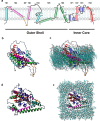
Structural model of prestin. a, schematic representation of the three-dimensional structure of the SulTP domain of prestin. Cylinders represent TM helices, and loops are indicated by black lines. The major structural features (outer shell and the inner core) are indicated at the bottom of the panel. The color of the different TM helices, ICF, HP1, and HP2 correspond to the coloring scheme of b–e. Lipid molecules are represented by gray spheres and lines. At the top is the extracellular region. b, side view of the ribbon representation of the three-dimensional structure of the SulTP domain of prestin. At the top is the extracellular region. N and C indicate the N- and C-terminal ends, respectively, of prestin. The ICF is yellow-colored. c, side view of 1-palmitoyl-2-oleoylphosphatidylcholine lipid bilayer-embedded prestin. The top is the extracellular region. d, extracellular view of prestin. e, extracellular view of 1-palmitoyl-2-oleoylphosphatidylcholine lipid bilayer-embedded prestin.
FIGURE 2.
FIGURE 2.
Molecular dynamics characterization of prestin. a, evolution of r.m.s.d. of the backbone atoms from the starting structure during three independent MD simulations. Simulation 1, black; simulation 2, green; simulation 3, red. b, root mean square fluctuation (RMSF) of residue positions. Major structural elements are designated by red bars on the upper y axis. c, tube representation of the central structure of the largest cluster of simulation 1. Regions of the inner core that form the proposed tunnel are color-coded as follows: HP1, yellow; TM7, dark gray; TM8, pink; ICF, green. The β-hairpin structure is indicated by arrows. N and C indicate the N- and C-terminal ends, respectively, of prestin. d and e, close-up of the tunnel in side- and top-down views, respectively. The color codes used are the same as in c. Interacting residues of the tunnel are shown in stick and indicated by black arrows.
FIGURE 3.
FIGURE 3.
Change in secondary structure during MD simulation of the structure of prestin. Secondary structure content was determined using the Dictionary of Secondary Structure of Proteins (DSSP) method.
FIGURE 4.
FIGURE 4.
The ECD spectra of 50 μm peptide fragments in different solutions. PTM8 (red), residues Ser478–Arg502 of prestin; GTM8 (black), residues Gly388–Glu418 of GltPh; ICF (blue), residues Ser133–Ile156 of prestin. deg, degrees.
FIGURE 5.
FIGURE 5.
Ionic interactions between positively and negatively charged side chains. a, the distance between center of masses of either the -ϵNH3+ group of lysine or -δNHC(NH2)2+ group of arginine and the -γCOO of glutamate during the MD simulation. Ionic interactions were considered when the distances between two oppositely charged groups were ≤0.5 nm. b, interacting residues in a are in stick and indicated by arrows in the tube representation of the central structure of the largest cluster of simulation 1.
FIGURE 6.
FIGURE 6.
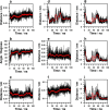
Interactions between various functional groups of amino acid residues in prestin during MD simulation. The interactions were assigned when distances and angles were in agreement with reported values in the literature (see Refs. 52, 53). Red lines are running averages at every 5-ns interval. a, distance between center of masses of the -δNHC(NH2)2+ group of Arg399 and the -βCOO of Asp485. b, angle between the planes of aromatic rings of Tyr367 and Tyr486; planes of the aromatic rings were assigned as a plane formed by the Cγ, Cϵ1, and Cϵ2 atoms. c, distance between the center of mass of the aromatic rings of Tyr367 and Tyr486. d, distance between center of masses of the aromatic ring of Tyr508 and the -δCH2 group of Leu142. e, distance between center of masses of the aromatic ring of Tyr501 and the peptide bond between Gly379 and Ile380. f, distance between center of masses of the aromatic ring of Tyr486 and the -δCH2 group of Arg399. g, distance between center of masses of the aromatic ring of Tyr508 and the -δCH2 group of Leu151. h, distance between center of masses of the aromatic ring of Tyr508 and the -γ1CH3 and -γ2CH3 groups of Val149. i, distance between center of masses of the aromatic ring of Tyr508 and the -δCH3 group of Ile144.
FIGURE 7.
FIGURE 7.
Structure of the chloride sensor of prestin. a, the backbone structure of the sensor is shown in gray ribbon, and the individual residues of HP1, TM7, TM8, and ICF that make up the chloride sensor are shown in stick. The red arrow indicates a possible path of chloride ions in the tunnel. b, stick representation of residues at the top of the channel. Dashed lines indicate the interactions between side chains of the following residues: Asp485-Arg399, ionic interaction; δCH2 of Arg399-aromatic side chain of Tyr486, CH-π interaction; aromatic side chain of Tyr486-aromatic side chain of Tyr367, π-π interaction. The gray sphere indicates a possible location of the Cl ion in the binding site.
FIGURE 8.
FIGURE 8.
NLC measured from HEK cells transfected with gPres and Tyr367 and Tyr486 mutant prestins. a, mean ± S.D. (error bars) of capacitance-voltage responses of gPres (WT) and mutant prestin without ICF (ICF del). NLC was normalized by Clin. Means ± S.D. of V1/2 and z values obtained from curve fitting the capacitance-voltage responses with the Boltzmann function are presented in the bottom panels. The confocal images in the top panels show membrane expression of WT and ICF-truncated proteins. b, mean ± S.D. (error bars) of capacitance-voltage responses of gPres (WT) and prestins with Y367A, Y367R, Y367F, Y486A, Y486R, and Y486F mutations. Means ± S.D. of V1/2 and z values (n = 11, 10, 12, 11, 11, and 12 for Y367A, Y367R, Y367F, Y486A, Y486R, and Y486F, respectively) obtained from curve fitting the capacitance-voltage responses with the Boltzmann function are presented in the bottom panels. Examples of membrane expression of the mutant proteins are presented in the top panels. From the left to right are Y367A, Y367R, Y367F, Y486A, Y486R, and Y486F. Scale bar, 5 μm.
FIGURE 9.
FIGURE 9.
NLC measured from HEK cells transfected with gPres and Tyr501 and Tyr508 mutant prestins. a, mean ± S.D. (error bars) of capacitance-voltage responses measured from prestin with Y501A, Y501R, Y501F, Y501W, Y508A, Y508R, Y508F, and Y508W mutations. Examples of confocal images of membrane expression of the mutant proteins are presented (from the left to right, Y501A, Y501R, Y501F, Y501W, Y508A, Y508R, Y508F, and Y508W). Capacitance-voltage responses from gPres (WT) were also plotted for comparison. b, means ± S.D. (error bars) of V1/2 and z values obtained from the capacitance-voltage responses fitted with the Boltzmann function. n = 11 (gPres), 10 (Y501A), 12 (Y501R), 11 (Y501W), 12 (Y501F), 10 (Y508A), 12 (Y508R), 11 (Y508W), 12 (Y508F), 11 (Y508W), and 12 (Y508F). c, NLC measured from double mutations at Tyr501 and Tyr508. Means ± S.D. (error bars) of V1/2 and z values were obtained from curve fitting with the Boltzmann function. n = 10 (Y501W/Y508W) and 12 (Y501F/Y508F). Examples of membrane expression of double mutant proteins are presented. Scale bar (applied to all images), 5 μm.
FIGURE 10.
FIGURE 10.
Proposed mechanism mediates unique function of prestin. Prestin at resting condition has binding sites for Cl ions through anion-π interaction. This interaction is much weaker than most of the other non-bonding interactions. The Cl ion can easily dissociate from the binding site and generate ion movement during voltage or mechanical stimulation. Binding or disruption of the interactions triggers ion movement (gating current) and subsequent conformational change. The locations of the four tyrosyl residues are indicated by the red circles. The sites for Arg399 and Asp485 and shown by the gray circles. The structure of prestin is represented by a shell indicated by the blue color-graded box, and the core is demarcated by yellow.

References

    1. Dallos P., Wu X., Cheatham M. A., Gao J., Zheng J., Anderson C. T., Jia S., Wang X., Cheng W. H., Sengupta S., He D. Z., Zuo J. (2008), Prestin-based outer hair cell motility is necessary for mammalian cochlear amplification. Neuron 58, 333–339 - PMC - PubMed
    1. Liberman M. C., Gao J., He D. Z., Wu X., Jia S., Zuo J. (2002) Prestin is required for outer hair cell motility and the cochlear amplifier. Nature 419, 300–304 - PubMed
    1. Fisher J. A., Nin F., Reichenbach T., Uthaiah R. C., Hudspeth A. J (2012) The spatial pattern of cochlear amplification. Neuron 76, 989–997 - PMC - PubMed
    1. Brownell W. E., Bader C. R., Bertrand D., de Ribaupierre Y. (1985) Evoked mechanical responses of isolated cochlear outer hair cells. Science 227, 194–196 - PubMed
    1. Zheng J., Shen W., He D. Z., Long K. B., Madison L. D., Dallos P. (2000) Prestin is the motor protein of cochlear outer hair cells. Nature 405, 149–155 - PubMed

Publication types

MeSH terms