Comparative Study
. 2015 Jul 31:21:811-27.
eCollection 2015.

Variable phenotypic expressivity in inbred retinal degeneration mouse lines: A comparative study of C3H/HeOu and FVB/N rd1 mice

Affiliations
Comparative Study

Variable phenotypic expressivity in inbred retinal degeneration mouse lines: A comparative study of C3H/HeOu and FVB/N rd1 mice

Michiel van Wyk et al. Mol Vis. .

Abstract

Purpose: Recent advances in optogenetics and gene therapy have led to promising new treatment strategies for blindness caused by retinal photoreceptor loss. Preclinical studies often rely on the retinal degeneration 1 (rd1 or Pde6b(rd1)) retinitis pigmentosa (RP) mouse model. The rd1 founder mutation is present in more than 100 actively used mouse lines. Since secondary genetic traits are well-known to modify the phenotypic progression of photoreceptor degeneration in animal models and human patients with RP, negligence of the genetic background in the rd1 mouse model is unwarranted. Moreover, the success of various potential therapies, including optogenetic gene therapy and prosthetic implants, depends on the progress of retinal degeneration, which might differ between rd1 mice. To examine the prospect of phenotypic expressivity in the rd1 mouse model, we compared the progress of retinal degeneration in two common rd1 lines, C3H/HeOu and FVB/N.

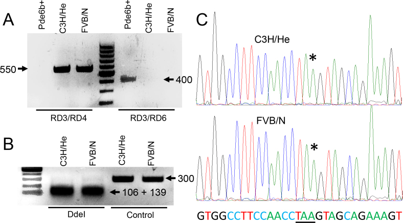
Methods: We followed retinal degeneration over 24 weeks in FVB/N, C3H/HeOu, and congenic Pde6b(+) seeing mouse lines, using a range of experimental techniques including extracellular recordings from retinal ganglion cells, PCR quantification of cone opsin and Pde6b transcripts, in vivo flash electroretinogram (ERG), and behavioral optokinetic reflex (OKR) recordings.

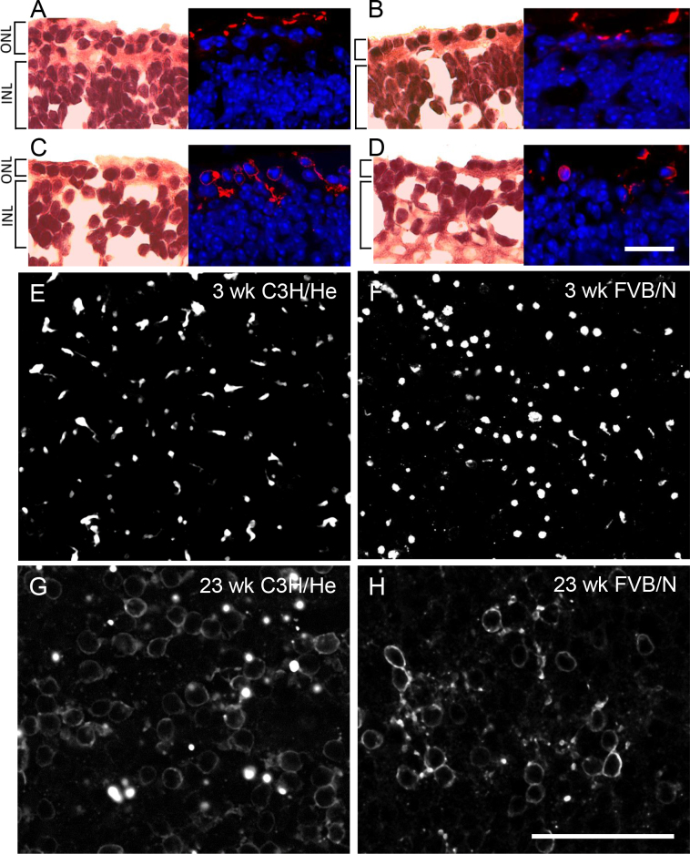
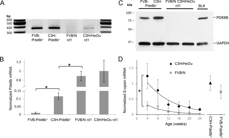
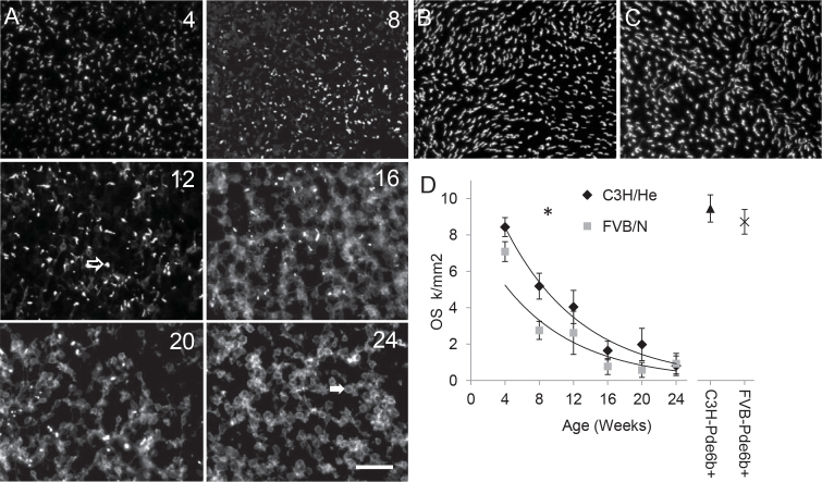
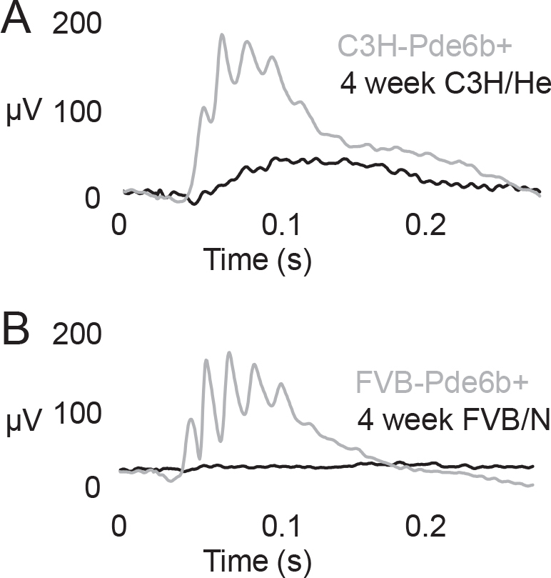
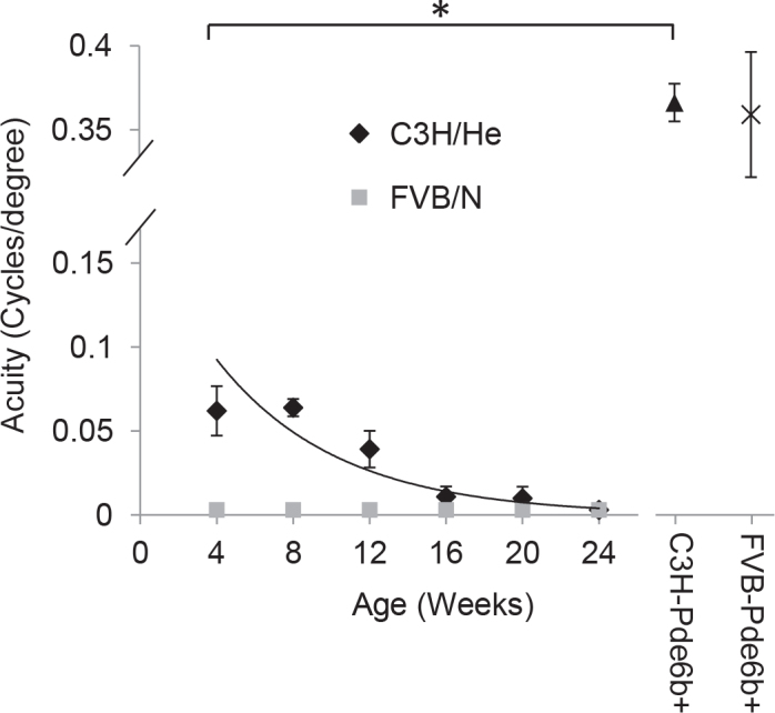
Results: We demonstrated a substantial difference in the speed of retinal degeneration and accompanying loss of visual function between the two rd1 lines. Photoreceptor degeneration and loss of vision were faster with an earlier onset in the FVB/N mice compared to C3H/HeOu mice, whereas the performance of the Pde6b(+) mice did not differ significantly in any of the tests. By postnatal week 4, the FVB/N mice expressed significantly less cone opsin and Pde6b mRNA and had neither ERG nor OKR responses. At 12 weeks of age, the retinal ganglion cells of the FVB/N mice had lost all light responses. In contrast, 4-week-old C3H/HeOu mice still had ERG and OKR responses, and we still recorded light responses from C3H/HeOu retinal ganglion cells until the age of 24 weeks. These results show that genetic background plays an important role in the rd1 mouse pathology.

Conclusions: Analogous to human RP, the mouse genetic background strongly influences the rd1 phenotype. Thus, different rd1 mouse lines may follow different timelines of retinal degeneration, making exact knowledge of genetic background imperative in all studies that use rd1 models.


Figures

Figure 1
Figure 1
Comparison of retinal morphologies of 3-week-old and 23-week-old FVB/N and C3H/HeOu mice. A–D: Longitudinal retinal cryosections stained with hematoxylin and eosin (left panels) and against S-opsin and 4',6-diamidino-2-phenylindole (DAPI) (right panels). EH: Retinal whole mounts stained against S-opsin. The outer nuclear layer (ONL) is two to three cell layers thick in 3-week-old C3H/HeOu mice (A), whereas the ONL of the FVB/N mice had only one layer of remnant photoreceptors (B). In 23-week mice of both lines, the ONL was one cell layer thick (C,D). At 3 weeks of age, the OS are more ordered and cylindrical in the C3H/HeOu retinas (A,E) compared to the FVB/N retinas (B,F). At 23 weeks (G,H), the OS are lost, and S-opsin is located in the cell bodies. Note that the FVB/N retina contains markedly fewer cones. Scale bars, (AD) 20 μm, (EH) 50 μm.
Figure 2
Figure 2
Confirmation of the rd1 genotype of FVB/N and C3H/HeOu mice. A: Presence of the provirus insertion in intron 1 of the PDE6B gene tested with Giménez and Montoliu’s three-primer method [54]. The primer pair RD3/RD4 amplifies a 550 bp product only in rd1 lines, whereas the primer pair RD3/RD6 amplifies a 400 bp product only in Pde6b+ control mice. B: The nonsense mutation in codon 347 of rd1 creates a DdeI restriction site. Digestion of a 300 bp PCR product spanning the mutation site with DdeI yielded two diagnostic fragments of 106 bp and 139 bp in both rd1 strains. Control: uncut 300 bp PCR product. C: DNA sequencing over Tyr347 (underlined) confirms the C→A mutation (*) introducing a stop codon (TAA) in both rd1 strains.
Figure 3
Figure 3
Quantification of Pde6b and S-opsin expression in 3-week old mice. A: Agarose gel electrophoresis of the qPCR products with Pde6b specific primers. The 359 bp Pde6b amplicon is detectable in all four mouse strains, however, at markedly different expression levels. B: Normalized Pde6b mRNA levels are significantly higher in C3H/HeOu compared to FVB/N mice, but indistinguishable in the two Pde6b+ control strains. For each group, n=7–8 retinas from four mice. C: β-PDE immunoreactivity of retinal protein extracts from Pde6b+, rd1, and C57BL/6 wild-type mice at postnatal day 21 using an antibody specific for the N-terminal region of β-PDE. An anti-GAPDH antibody detecting a band of approximately 37 kDa was used as a loading control. No β-PDE expression is detected in rd1 strains. D: Age-dependent S-opsin transcript levels. No significant differences exist between the two Pde6b+ lines and young, 4- to 8-week-old C3H/HeOu mice. In contrast, the S-opsin transcripts in the retinas from 4-week-old FVB/N mice were significantly lower than in coeval C3H/HeOu retinas. For each group, n=6 retinas from three mice. Error bars indicate standard deviation (SD). The fits are exponential with the R-square values for C3H/HeOu and FVB/N at 0.92 and 0.87, respectively.
Figure 4
Figure 4
Immunocytochemical detection of cone opsin. Images were always taken from the retinal area with the highest density of labeled cones, typically in the mid-periphery of the retina. A: Examples of S-opsin staining in C3H/HeOu retinas demonstrate the progressive loss of cone outer segments and a concomitant accumulation of S-opsin in the cell bodies. One misshaped outer segment (clear arrow) and one soma without outer segments (OS) are indicated (solid arrow). Age, in weeks, is indicated in the top right of every panel. Scale bar=50 µm. B, C: S-opsin staining in wild-type C3H-Pde6b+ (B) and FVB-Pde6b+ (C) retinas were similar. D: Summary graph of OS counts in rd1 C3H/HeOu and FVB/N mice relative to the Pde6b+ mouse lines. For each group, n=8 retinas from four mice. The fits are exponential with the R-square values for C3H/HeOu and FVB/N at 0.94 and 0.8, respectively.
Figure 5
Figure 5
Fraction of retinal ganglion cells responding to a light step in isolated retinas. In both seeing Pde6b+ mouse lines, over 90% of the retinal ganglion cells (GCs) responded to light, with no significant difference between the C3H/He-Pde6b+ and FVB/N-Pde6b+ lines. At 4 weeks, the fraction of responding GCs in the C3H/HeOu mice approximated that of Pde6b+ mice, while the number of responding cells in the age-matched FVB/N mice was already significantly reduced. No responding GCs remained in the FVB/N mice at 12 weeks of age compared to the C3H/HeOu mice where retinal responsiveness disappeared only at 24 weeks of age. For each group, n=6 retinas from six different mice.
Figure 6
Figure 6
Photopic flash electroretinogram (ERG) recordings. In both Pde6b+ mouse lines, we recorded strong responses, with no significant difference in amplitudes. In contrast, the ERGs recorded from the 4-week-old C3H/HeOu mice were markedly weaker with a delayed b-wave that disappeared in older mice. No ERG response was recorded in the FVB/N mice at any time point. For each group, n=6 mice.
Figure 7
Figure 7
Maximum spatial acuity of the optokinetic reflex (OKR) responses. Although both Pde6b+ mouse lines responded to spatial frequencies characteristic of wild-type mice with no significant difference between the two mouse lines, only young 4-week-old C3H/HeOu mice reliably responded with a tracking reflex and only to relatively low spatial frequencies (0.062±0.02 cyc/deg). The number of C3H/HeOu mice with an OKR response decreased steadily with age and approached zero at 24 weeks. The FVB/N mice did not have a detectable OKR at any age. The fit is exponential with an R-square value of 0.92.

References

    1. Busskamp V, Roska B. Optogenetic approaches to restoring visual function in retinitis pigmentosa. Curr Opin Neurobiol. 2011;21:942–6. - PubMed
    1. Cronin T, Vandenberghe L, Hantz P, Juttner J, Reimann A, Kacsó A, Huckfeldt R, Busskamp V, Kohler H, Lagali P, Roska B, Bennett J. Efficient transduction and optogenetic stimulation of retinal bipolar cells by a synthetic adeno-associated virus capsid and promoter. EMBO Mol Med. 2014;6:1175–90. - PMC - PubMed
    1. Caporale N, Kolstad K, Lee T, Tochitsky I, Dalkara D, Trauner D, Kramer R, Dan Y, Isacoff E, Flannery J. LiGluR restores visual responses in rodent models of inherited blindness. Mol Ther. 2011;19:1219–29. - PMC - PubMed
    1. Chang B, Hawes NL, Hurd RE, Davisson MT, Nusinowitz S, Heckenlively JR. Retinal degeneration mutants in the mouse. Vision Res. 2002;42:517–25. - PubMed
    1. Bowes C, Li T, Frankel WN, Danciger M, Coffin JM, Applebury ML, Farber DB. Localization of a retroviral element within the rd gene coding for the beta subunit of cGMP phosphodiesterase. Proc Natl Acad Sci USA. 1993;90:2955–9. - PMC - PubMed

Publication types

MeSH terms