Case Reports
. 2015 Dec;167A(12):2975-84.
doi: 10.1002/ajmg.a.37297. Epub 2015 Aug 18.

Exome analysis of a family with Wolff-Parkinson-White syndrome identifies a novel disease locus

Affiliations
Case Reports

Exome analysis of a family with Wolff-Parkinson-White syndrome identifies a novel disease locus

Neil E Bowles et al. Am J Med Genet A. 2015 Dec.

Abstract

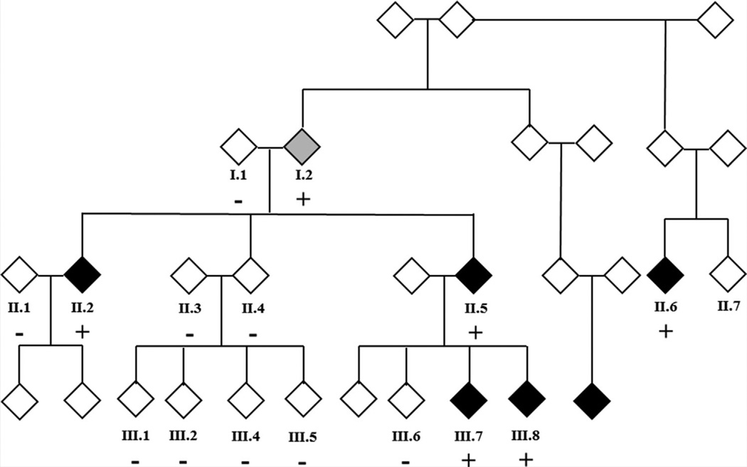
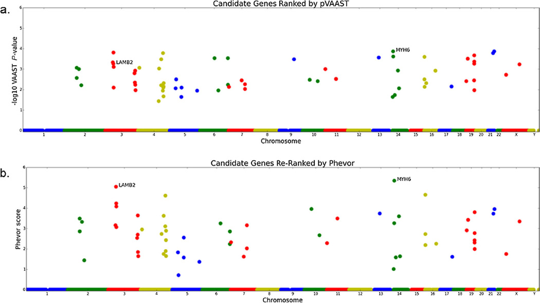
Wolff-Parkinson-White (WPW) syndrome is a common cause of supraventricular tachycardia that carries a risk of sudden cardiac death. To date, mutations in only one gene, PRKAG2, which encodes the 5'-AMP-activated protein kinase subunit γ-2, have been identified as causative for WPW. DNA samples from five members of a family with WPW were analyzed by exome sequencing. We applied recently designed prioritization strategies (VAAST/pedigree VAAST) coupled with an ontology-based algorithm (Phevor) that reduced the number of potentially damaging variants to 10: a variant in KCNE2 previously associated with Long QT syndrome was also identified. Of these 11 variants, only MYH6 p.E1885K segregated with the WPW phenotype in all affected individuals and was absent in 10 unaffected family members. This variant was predicted to be damaging by in silico methods and is not present in the 1,000 genome and NHLBI exome sequencing project databases. Screening of a replication cohort of 47 unrelated WPW patients did not identify other likely causative variants in PRKAG2 or MYH6. MYH6 variants have been identified in patients with atrial septal defects, cardiomyopathies, and sick sinus syndrome. Our data highlight the pleiotropic nature of phenotypes associated with defects in this gene.

Keywords: MYH6; Wolff-Parkinson-White; whole exome sequencing.


Conflict of interest statement

Conflicts of interests: none.

Figures

FIG. 1
FIG. 1
Family K32326 pedigree. The gender of family members is masked for confidentiality. Black symbols represent patients with WPW and a gray symbol represents the patient with a diagnosis of SVT (Patient 1:2): autosomal dominant inheritance, with incomplete penetrance, is the most likely genetic model. Only participants in the study for whom DNA is available for analysis are numbered. +: Positive for MYH6 c.5653G>A, p.Glu1885Lys; −: Negative for MYH6 c.5653G>A, p.Glu1885Lys.
FIG. 2
FIG. 2
Representative ECGs from selected members of K32326. Top row: example of a normal resting ECG (left panel). Right panel, resting ECG from patient II-5 in sinus rhythm, showing classic short PR interval and ventricular pre-excitation or delta wave (arrow). Bottom row: ECG from patient III-7 shows short PR interval and subtle ventricular pre-excitation (left panel). ECG from patient III-8 reveals markedly short PR interval and prominent ventricular pre-excitation (right panel).
FIG. 3
FIG. 3
Manhattan plots of the pVAAST and Phevor results. A: Manhattan plot of pVAAST scores for all protein-coding genes in Human Genome release hg19; each dot is a single gene. B: Manhattan plot of Phevor scores obtained by using the pVAAST results in conjunction with the phenotype terms describing symptoms of Wolff–Parkinson–White. The x-axis shows the genomic location of each gene arranged by position along the chromosomes. The y-axis is the Phevor or pVAAST score. MYH6 and LAMB2 are highlighted in each plot as they are the highest scoring genes by the combination of analyses.
FIG. 4
FIG. 4
Conservation analysis of MYH6. Alignments of MYH6 across several species of the amino acids surrounding Glutamic acid 1885 generated using the PRALINE multiple sequence alignment tool (http://www.ibi.vu.nl/programs/pralinewww/). [Heringa, 1999] The scoring scheme is from 0 for the least conserved alignment position, up to 10 for the most conserved alignment position (represented by *) and the color assignments for each score are shown at the bottom. Note the complete conservation of this amino acid and the high degree of conservation across the domain.

References

    1. Abbott GW, Sesti F, Splawski I, Buck ME, Lehmann MH, Timothy KW, Keating MT, Goldstein SA. MiRP1 forms IKr potassium channels with HERG and is associated with cardiac arrhythmia. Cell. 1999;97:175–187. - PubMed
    1. Adzhubei IA, Schmidt S, Peshkin L, Ramensky VE, Gerasimova A, Bork P, Kondrashov AS, Sunyaev SR. A method and server for predicting damaging missense mutations. Nat Methods. 2010;7:248–249. - PMC - PubMed
    1. Aggarwal P, Gill-Randall R, Wheatley T, Buchalter MB, Metcalfe J, Alcolado JC. Identification of mtDNA mutation in a pedigree with gestational diabetes, deafness, Wolff–Parkinson–White syndrome and placenta accreta. Hum Hered. 2001;51:114–116. - PubMed
    1. Arrington CB, Bleyl SB, Matsunami N, Bonnell GD, Otterud BE, Nielsen DC, Stevens J, Levy S, Leppert MF, Bowles NE. Exome analysis of a family with pleiotropic congenital heart disease. Circ Cardiovasc Genet. 2012;5:175–182. - PMC - PubMed
    1. Arrington CB, Sower CT, Chuckwuk N, Stevens J, Leppert MF, Yetman AT, Bowles NE. Absence of TGFBR1 and TGFBR2 mutations in patients with bicuspid aortic valve and aortic dilation. Am J Cardiol. 2008;102:629–631. - PubMed

Publication types

LinkOut - more resources