

Flow Cytofluorimetric Analysis of Anti-LRP4 (LDL Receptor-Related Protein 4) Autoantibodies in Italian Patients with Myasthenia Gravis
- PMID: 26284792
- PMCID: PMC4540439
- DOI: 10.1371/journal.pone.0135378
Flow Cytofluorimetric Analysis of Anti-LRP4 (LDL Receptor-Related Protein 4) Autoantibodies in Italian Patients with Myasthenia Gravis
Abstract
Background: Myasthenia gravis (MG) is an autoimmune disease in which 90% of patients have autoantibodies against the muscle nicotinic acetylcholine receptor (AChR), while autoantibodies to muscle-specific tyrosine kinase (MuSK) have been detected in half (5%) of the remaining 10%. Recently, the low-density lipoprotein receptor-related protein 4 (LRP4), identified as the agrin receptor, has been recognized as a third autoimmune target in a significant portion of the double sero-negative (dSN) myasthenic individuals, with variable frequency depending on different methods and origin countries of the tested population. There is also convincing experimental evidence that anti-LRP4 autoantibodies may cause MG.
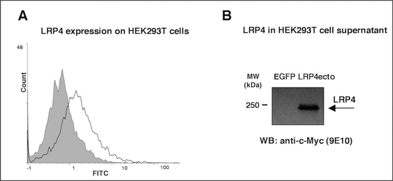
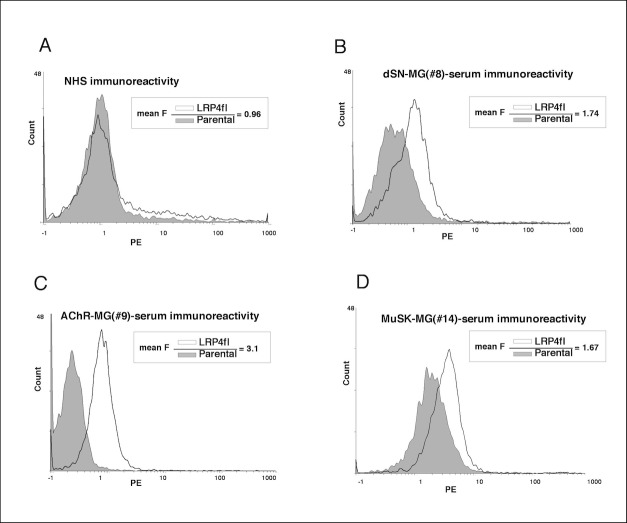
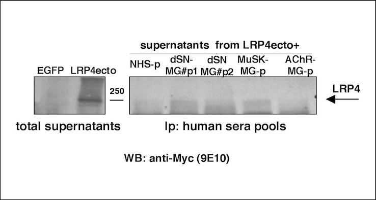
Methods: The aim of this study was to test the presence and diagnostic significance of anti-LRP4 autoantibodies in an Italian population of 101 myasthenic patients (55 dSN, 23 AChR positive and 23 MuSK positive), 45 healthy blood donors and 40 patients with other neurological diseases as controls. All sera were analyzed by a cell-based antigen assay employing LRP4-transfected HEK293T cells, along with a flow cytofluorimetric detection system.
Results: We found a 14.5% (8/55) frequency of positivity in the dSN-MG group and a 13% frequency of co-occurrence (3/23) in both AChR and MuSK positive patients; moreover, we report a younger female prevalence with a mild form of disease in LRP4-positive dSN-MG individuals.
Conclusion: Our data confirm LRP4 as a new autoimmune target, supporting the value of including anti-LRP4 antibodies in further studies on Myasthenia gravis.
Conflict of interest statement
Figures
References
-
- Lindstrom J (1977) An assay for antibodies to human acetylcholine receptor in serum from patients with myasthenia gravis. Clin Immunol Immunopathol 7: 36–43. - PubMed
-
- Hoch W, McConville J, Helms S, Newsom-Davis J, Melms A, Vincent A (2001) Auto-antibodies to the receptor tyrosine kinase MuSK in patients with myasthenia gravis without acetylcholine receptor antibodies. Nat Med 7: 365–368. - PubMed
Publication types
MeSH terms
Substances
LinkOut - more resources
Full Text Sources
Other Literature Sources
Medical
Miscellaneous
