Case Reports
. 2015 Dec;173(6):1505-13.
doi: 10.1111/bjd.14073. Epub 2015 Oct 29.

Unusual cutaneous features associated with a heterozygous gain-of-function mutation in IFIH1: overlap between Aicardi-Goutières and Singleton-Merten syndromes

Affiliations
Case Reports

Unusual cutaneous features associated with a heterozygous gain-of-function mutation in IFIH1: overlap between Aicardi-Goutières and Singleton-Merten syndromes

A-C Bursztejn et al. Br J Dermatol. 2015 Dec.

Abstract

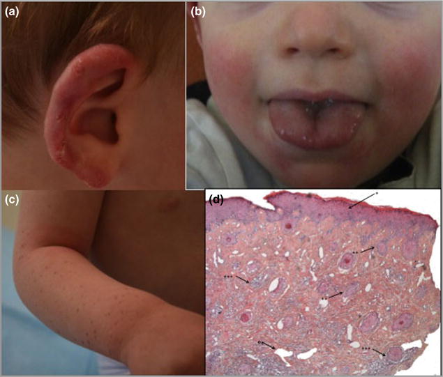
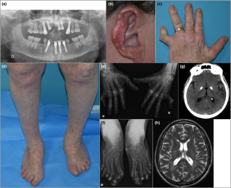
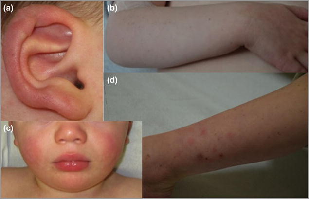
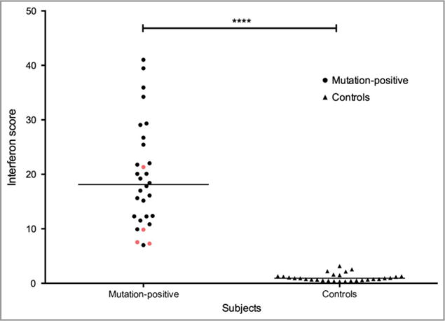
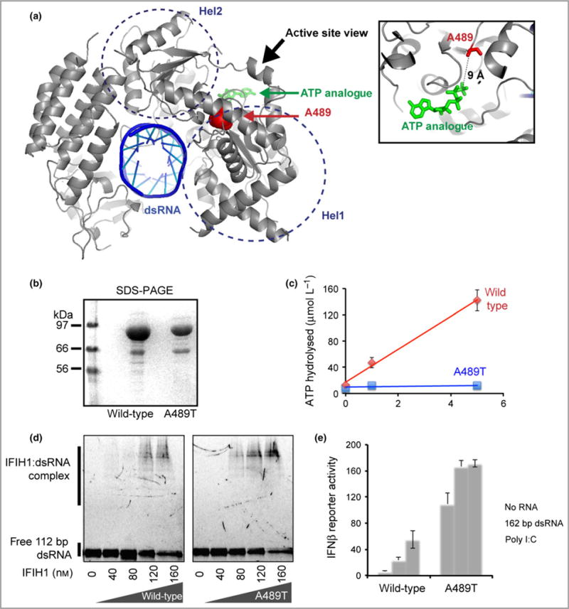
Cutaneous lesions described as chilblain lupus occur in the context of familial chilblain lupus or Aicardi-Goutières syndrome. To date, seven genes related to Aicardi-Goutières syndrome have been described. The most recently described encodes the cytosolic double-stranded RNA receptor IFIH1 (also known as MDA5), a key component of the antiviral type I interferon-mediated innate immune response. Enhanced type I interferon signalling secondary to gain-of-function mutations in IFIH1 can result in a range of neuroinflammatory phenotypes including classical Aicardi-Goutières syndrome. It is of note that none of the patients with a neurological phenotype so far described with mutations in this gene was reported to demonstrate cutaneous involvement. We present a family segregating a heterozygous pathogenic mutation in IFIH1 showing dermatological involvement as a prominent feature, variably associated with neurological disturbance and premature tooth loss. All three affected individuals exhibited increased expression of interferon-stimulated genes in whole blood, and the mutant protein resulted in enhanced interferon signalling in vitro, both in the basal state and following ligand stimulation. Our results further extend the phenotypic spectrum associated with mutations in IFIH1, indicating that the disease can be confined predominantly to the skin, while also highlighting phenotypic overlap with both Aicardi-Goutières syndrome and Singleton-Merten syndrome.


Conflict of interest statement

Conflicts of interest

None declared.

Figures

Fig 1
Fig 1
Clinical images of the proband. (a) Erythema and ulceration of the outer helix of the right ear. (b) Erythema of the cheeks bilaterally. No lesions of the tongue were noticed. (c) Lentigines of the right forearm. (d) Histology of the ear helix demonstrating slight acanthosis of the epidermis (*), interface dermatitis located mostly in the follicles (**) and superficial and deep infiltrate along the adnexae and around the vessels (***). In addition most capillaries are dilated (°) (magnification level, d:HES × 10).
Fig 2
Fig 2
Clinical images of the 41-year-old father. (a) Pantomogram showing loss of permanent teeth. The remaining dentition, molars and premolars demonstrate root hypoplasia. (b) Scarring and tissue loss of the outer ear helix following chronic ulceration. (c) Deformities of the metacarpophalangeal and interphalangeal joints of the left hand, and multiple lentigines. (d) Hallux valgus, psoriatic lesions over the knees and multiple lentigines. (e) Radiography showing joint deformities on the hands. (f) Radiography showing joint deformities on the feet. (g) Brain imaging at age 41 years showing bilateral calcification of the globus pallidus on computed tomography. (h) Brain imaging at age 41 years illustrating high signal in the white matter around the posterior poles of the lateral ventricles on axial T2-weighted magnetic resonance imaging.
Fig 3
Fig 3
Clinical images of the younger sibling of the proband. (a) Erythema and ulceration of the outer helix of the right ear. (b) Multiple lentigines of the right forearm. (c) Bilateral erythema of the cheeks. (d) Lower-limb atrophic scars, which occurred following resolution of papular lesions.
Fig 4
Fig 4
Individuals with the p.Ala489Thr interferon-induced helicase C domain-containing protein 1 (IFIH1) mutant demonstrate elevated levels of interferon-stimulated genes compared with controls. An interferon score was calculated from the median fold change in RQ value for a panel of six interferon signature genes measured using quantitative polymerase chain reaction – IFI27 (Hs01086370_m1), IFI44L (Hs00199115_m1), IFIT1 (Hs00356631_g1), ISG15 (Hs00192713_m1), RSAD2 (Hs01057264_m1) and SIGLEC1 (Hs00988063_m1) – normalized to the expression levels of HPRT1 (Hs03929096_g1) and 18S RNA (Hs999999001_s1). All three affected members of the family (red) display a score that is comparable with previously reported cases (black) with IFIH1-related disease, and is significantly higher than in control samples (****P < 0.0001). Levels were measured on two separate occasions in one child in the reported family.
Fig 5
Fig 5
The p.Ala489Thr interferon-induced helicase C domain-containing protein 1 (IFIH1) mutant confers a gain of interferon signalling. (a) Mapping of the altered residue A489 (red sphere) onto the structure of IFIH1 Δ2CARD (grey) bound by double-stranded (ds)RNA (blue) and ATP analogue (green) (Protein Data Bank 4GL2). (b) Sodium dodecylsulfate polyacrylamide gel electrophoresis (SDS-PAGE) analysis (Coomassie stain) of the purified wild-type and mutant IFIH1. (c) ATP hydrolysis activity (mean ± SD, n = 3 biological replicates) of wild-type and mutant IFIH1. (d) Electrophoretic mobility shift assay of purified wild-type and mutant IFIH1 with 112-bp dsRNA. The gel image is representative of three independent experiments in the presence of ATP. (e) Interferon (IFN)-β reporter activity (mean ± SD, n = 3 biological replicates) of Flag-tagged wild-type and mutant IFIH1 with and without stimulation with 162-bp dsRNA or polyinosinic–polycytidylic acid (poly I:C) in HEK293T cells. The results are representative of three independent experiments.

Comment in

References

    1. Crow YJ, Chase DS, Lowenstein Schmidt J, et al. Characterization of human disease phenotypes associated with mutations in TREX1, RNASEH2A, RNASEH2B, RNASEH2C, SAMHD1, ADAR, and IFIH1. Am J Med Genet A. 2015;167A:296–312. - PMC - PubMed
    1. Rice GI, del Toro Duany Y, Jenkinson EM, et al. Gain-of-function mutations in IFIH1 cause a spectrum of human disease phenotypes associated with upregulated type I interferon signaling. Nat Genet. 2014;46:503–9. - PMC - PubMed
    1. Oda H, Nakagawa K, Abe J, et al. Aicardi-Goutières syndrome is caused by IFIH1 mutations. Am J Hum Genet. 2014;95:121–5. - PMC - PubMed
    1. Rice G, Newman WG, Dean J, et al. Heterozygous mutations in TREX1 cause familial chilblain lupus and dominant Aicardi-Goutières syndrome. Am J Hum Genet. 2007;80:811–15. - PMC - PubMed
    1. Günther C, Meurer M, Stein A, et al. Familial chilblain lupus – a monogenic form of cutaneous lupus erythematosus due to a heterozygous mutation in TREX1. Dermatology. 2009;219:162–6. - PubMed

Publication types

MeSH terms

Substances

Supplementary concepts