. 2016 Jan 1;39(1):155-9.
doi: 10.5665/sleep.5338.

Sleep-Dependent Potentiation in the Visual System Is at Odds with the Synaptic Homeostasis Hypothesis

Affiliations

Sleep-Dependent Potentiation in the Visual System Is at Odds with the Synaptic Homeostasis Hypothesis

Jaclyn Durkin et al. Sleep. .

Abstract

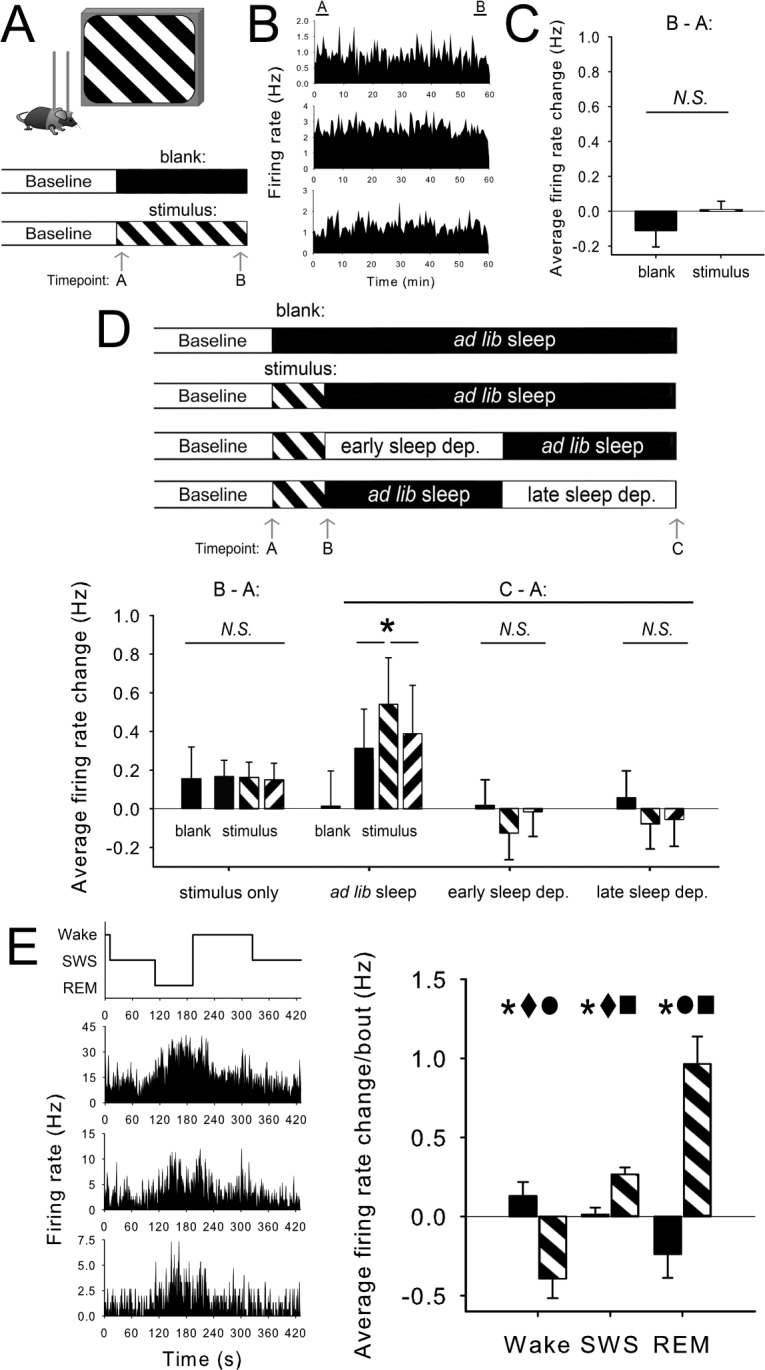
Study objectives: Two commentaries recently published in SLEEP came to very different conclusions regarding how data from a mouse model of sleep-dependent neural plasticity (orientation-specific response potentiation; OSRP) fit with the synaptic homeostasis hypothesis (SHY). To assess whether SHY offers an explanatory mechanism for OSRP, we present new data on how cortical neuron firing rates are modulated as a function of novel sensory experience and subsequent sleep in this model system.

Methods: We carried out longitudinal extracellular recordings of single-neuron activity in the primary visual cortex across a period of novel visual experience and subsequent sleep or sleep deprivation. Spontaneous neuronal firing rates and visual responses were recorded from the same population of visual cortex neurons before control (blank screen) or novel (oriented grating) stimulus presentation, immediately after stimulus presentation, and after a period of subsequent ad lib sleep or sleep deprivation.

Results: Firing rate responses to visual stimuli were unchanged across waking experience, regardless of whether a blank screen or an oriented grating stimulus was presented. Firing rate responses to stimuli of the presented stimulus orientation were selectively enhanced across post-stimulus sleep, but these changes were blocked by sleep deprivation. Neuronal firing increased significantly across bouts of post-stimulus rapid eye movement (REM) sleep and slow wave sleep (SWS), but not across bouts of wake.

Conclusions: The current data suggest that following novel visual experience, potentiation of a subset of V1 synapses occurs across periods of sleep. This finding cannot be explained parsimoniously by SHY.

Keywords: cerebral cortex; electrophysiology; sensory processing; synaptic plasticity.


Figures

Figure 1
Figure 1
Cortical neurons' firing rates do not change across waking visual experience which induces OSRP, but do increase across subsequent sleep. (A) To assess firing rate changes over waking experience, firing rates were compared during the first (Timepoint A) and last (Timepoint B) 5-min windows of oriented grating (stimulus) presentation or blank screen (blank) presentation, beginning at lights-on. (B) Firing rate histograms for three representative V1 neurons during 1-h stimulus presentation. (C) Firing rate changes for individual neurons (in Hz) were not significantly changed across stimulus presentation, and were not different between stimulus (n = 20 experiments [8 from a previous study], 268 neurons) and blank screen (n = 3 experiments, 44 neurons) conditions. (D) No differences in either spontaneous firing rate or orientation-specific responses (presented and orthogonal orientations are shown) were seen immediately after blank screen or stimulus presentation (Timepoint B). After subsequent ad lib sleep (Timepoint C; n = 11 experiments [4 from a previous study], 137 neurons), firing rate responses were selectively enhanced for the presented stimulus. *P < 0.05 for presented vs. orthogonal, presented vs. blank, Holm-Sidak post hoc test, P < 0.05, RM ANOVA. Post-sleep firing rate changes following blank screen presentation (n = 8 experiments [4 from a previous study], 105 neurons) were negligible. Subsets of mice underwent behavioral sleep deprivation in the first (early sleep dep., n = 14 experiments [4 from a previous study], 176 neurons) or second (late sleep dep., n = 13 experiments [3 from a previous study], 166 neurons) half of the post stimulus sleep period. In both sleep deprivation conditions, response rate changes across the day were negligible, and stimulus-specific potentiation of responses was lost. (E) To determine how neuronal firing changed during sleep and wake bouts, firing rates were averaged over the first and last 30 seconds of individual bouts of wake, SWS, or REM. Changes in firing were calculated for each bout ≥ 1 min over the first 4 h following presentation of oriented gratings (striped bars; n = 509, 1152, and 287 measurements from 4 mice for wake, SWS, and REM, respectively) or blank screen (black bars; n = 630, 1625, and 236 measurements from 4 mice). Bouts with zero firing were excluded from analysis. *P < 0.005 for stimulus vs. blank screen; ■, ●, and ♦ indicate P < 0.001 vs. wake, SWS, and REM, respectively in the stimulus condition, 2-way RM ANOVA with Holm-Sidak post hoc test.

References

    1. Heller C. The ups and downs of synapses during sleep and learning. Sleep. 2014;37:1157–8. - PMC - PubMed
    1. Cirelli C, Tononi G. Sleep and synaptic homeostasis. Sleep. 2015;38:161–2. - PMC - PubMed
    1. Aton SJ, Suresh A, Broussard C, Frank MG. Sleep promotes cortical response potentiation following visual experience. Sleep. 2014;37:1163–70. - PMC - PubMed
    1. Tononi G, Cirelli C. Sleep and the price of plasticity: from synaptic and cellular homeostasis to memory consolidation and integration. Neuron. 2014;81:12–34. - PMC - PubMed
    1. Cirelli C, Gutierrez CM, Tononi G. Extensive and divergent effects of sleep and wakefulness on brain gene expression. Neuron. 2004;41:35–43. - PubMed

Publication types