Review
. 2015 Aug 18;5(8):e623.
doi: 10.1038/tp.2015.115.

Schizophrenia: a tale of two critical periods for prefrontal cortical development

Affiliations
Review

Schizophrenia: a tale of two critical periods for prefrontal cortical development

L D Selemon et al. Transl Psychiatry. .

Abstract

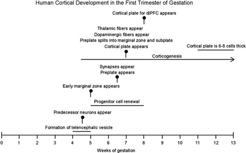
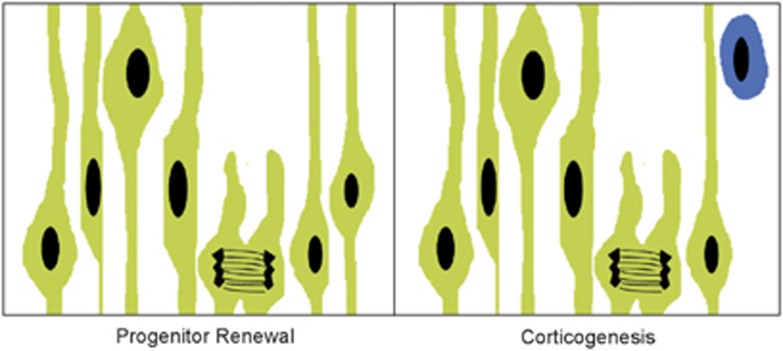
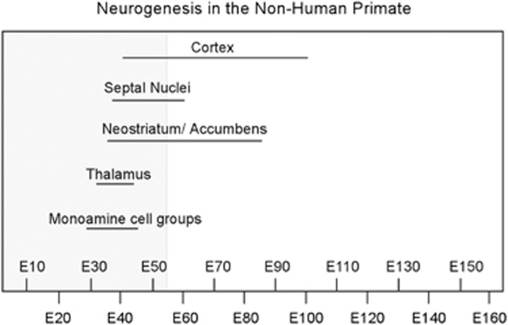
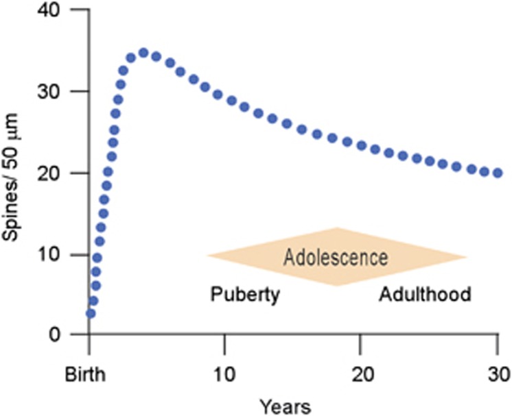
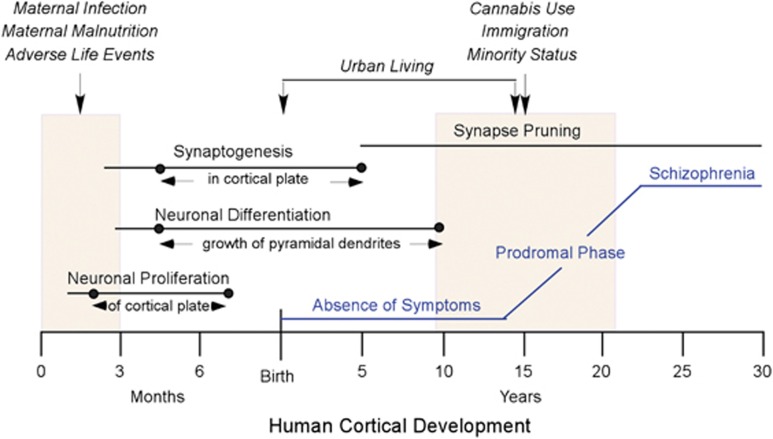
Schizophrenia is a disease of abnormal brain development. Considerable evidence now indicates that environmental factors have a causative role in schizophrenia. Elevated incidence of the disease has been linked to a wide range of disturbances in the prenatal environment and to social factors and drug intake during adolescence. Here we examine neurodevelopment of the prefrontal cortex in the first trimester of gestation and during adolescence to gain further insight into the neurodevelopmental processes that may be vulnerable in schizophrenia. Early embryonic development of the prefrontal cortex is characterized by cell proliferation, including renewal of progenitor cells, generation of early transient cell populations and neurogenesis of subcortical populations. Animal models show that curtailing early gestational cell proliferation produces schizophrenia-like pathology in the prefrontal cortex and mimics key behavioral and cognitive symptoms of the disease. At the other end of the spectrum, elimination of excitatory synapses is the fundamental process occurring during adolescent maturation in the prefrontal cortex. Adverse social situations that elevate stress increase dopamine stimulation of the mesocortical pathway and may lead to exaggerated synaptic pruning during adolescence. In a non-human primate model, dopamine hyperstimulation has been shown to decrease prefrontal pyramidal cell spine density and to be associated with profound cognitive dysfunction. Development of the prefrontal cortex in its earliest stage in gestation and in its final stage in adolescence represents two critical periods of vulnerability for schizophrenia in which cell proliferation and synaptic elimination, respectively, may be influenced by environmental factors.


Figures

Figure 1
Figure 1
Important milestones in human cortical development are indicated by vertical markers. Ongoing processes are shown as horizontal lines. Note that corticogenesis begins at ~4.5 gw with genesis of preplate neurons: first predecessor cells followed by Cajal–Retzius and other preplate neurons. Corticogenesis of neurons populating the cortical plate commences later, in the 8th gw for the dorsolateral prefrontal cortex (dlPFC). gw, gestational weeks.
Figure 2
Figure 2
Illustration of symmetric cell division (left) and asymmetric cell division (right) in the ventricular zone of the prospective telencephalon. Progenitor cells (green), also known as radial glial cells, have elongated cell processes that are attached to the ventricular and pial surfaces and form a pseudostratified epithelium. Cell nuclei move toward the pial surface during DNA synthesis and then toward the ventricular surface just before mitosis. In early phases of cortical development, symmetric division gives rise to two identical daughter cells that are both progenitor cells (progenitor renewal). Later in development, asymmetric division (corticogenesis) produces a progenitor cell and a post-mitotic neuron (blue).
Figure 3
Figure 3
Timeline showing the duration of neurogenesis for various brain structures in the non-human primate as a function of gestational age in embryonic days (E). Neurogenesis occurs in a staggered manner, beginning and ending earlier for subcortical neurons than for cortical neurons. Corticogenesis commences near the end of the first trimester (shaded area) and extends throughout midgestation. Although tritiated thymidine dating of neurogenesis is not possible in the human, morphologic evidence suggests that corticogenesis spans a similar time period in human gestation. Source: on the basis of data from Rakic and colleagues., , ,
Figure 4
Figure 4
Spine density (blue dotted line) on pyramidal cells in the human dorsolateral prefrontal cortex as a function of postnatal age. Note that there is a net spine loss, indicating pruning of excitatory synapses, beginning in childhood, continuing throughout adolescence, and extending into early adulthood. Adolescence is a period with imprecise boundaries beginning with the onset of puberty and ending at the point in which adult responsibilities are assumed. Source: on the basis of data from apical proximal oblique dendrites in Petanjek et al. and in agreement with Huttenlocher and Dabholkar.
Figure 5
Figure 5
Summary showing human cortical development, environmental factors that increase the risk for later development of schizophrenia and clinical progression of the disease. Major processes in prenatal cortical development (in months) and postnatal cortical development (in years) are indicated by horizontal lines. Dots on the lines indicate the extent of those processes as they occur in the cortex proper, as detailed in text between arrows. Shaded boxes indicate the first trimester of gestation and adolescence. Above the shaded boxes, environmental factors (in italics) that may have an impact on neural development and increase the risk of developing schizophrenia are shown (vertical arrows). The clinical course of schizophrenia from an asymptomatic state in childhood to full expression of the disease in late adolescence/early adulthood is shown in blue.

References

    1. Weinberger DR. Implications of normal brain development for the pathogenesis of schizophrenia. Arch Gen Psychiatry. 1987;44:660–669. - PubMed
    1. Johnstone EC, Crow TJ, Frith CD, Husband J, Kreel L. Cerebral ventricular size and cognitive impairment in chronic schizophrenia. Lancet. 1976;2:924–926. - PubMed
    1. Weinberger DR, Torrey EF, Neophytides AN, Wyatt RJ. Lateral cerebral ventricular enlargement in chronic schizophrenia. Arch Gen Psychiatry. 1979;36:735–739. - PubMed
    1. Feinberg I. Schizophrenia: caused by a fault in programmed synaptic elimination during adolescence. J Psychiatr Res. 1982;17:319–330. - PubMed
    1. Brown AS. Epidemiologic studies of exposure to prenatal infection and risk of schizophrenia and autism. Dev Neurobiol. 2012;72:1272–1276. - PMC - PubMed

Publication types