. 2015 Aug 18;10(8):e0135582.
doi: 10.1371/journal.pone.0135582. eCollection 2015.

High Inorganic Phosphate Intake Promotes Tumorigenesis at Early Stages in a Mouse Model of Lung Cancer

Affiliations

High Inorganic Phosphate Intake Promotes Tumorigenesis at Early Stages in a Mouse Model of Lung Cancer

Somin Lee et al. PLoS One. .

Abstract

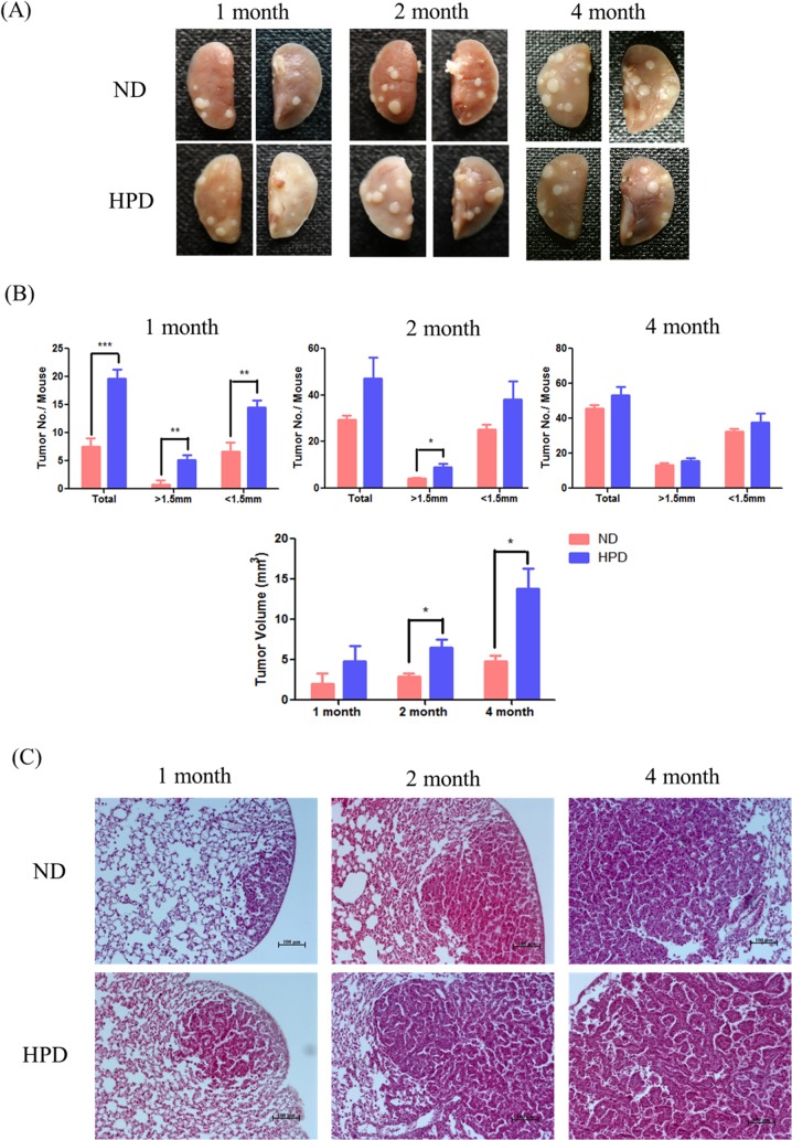
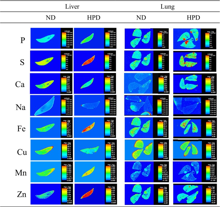
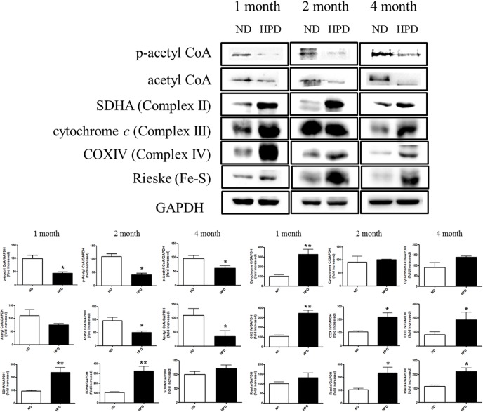
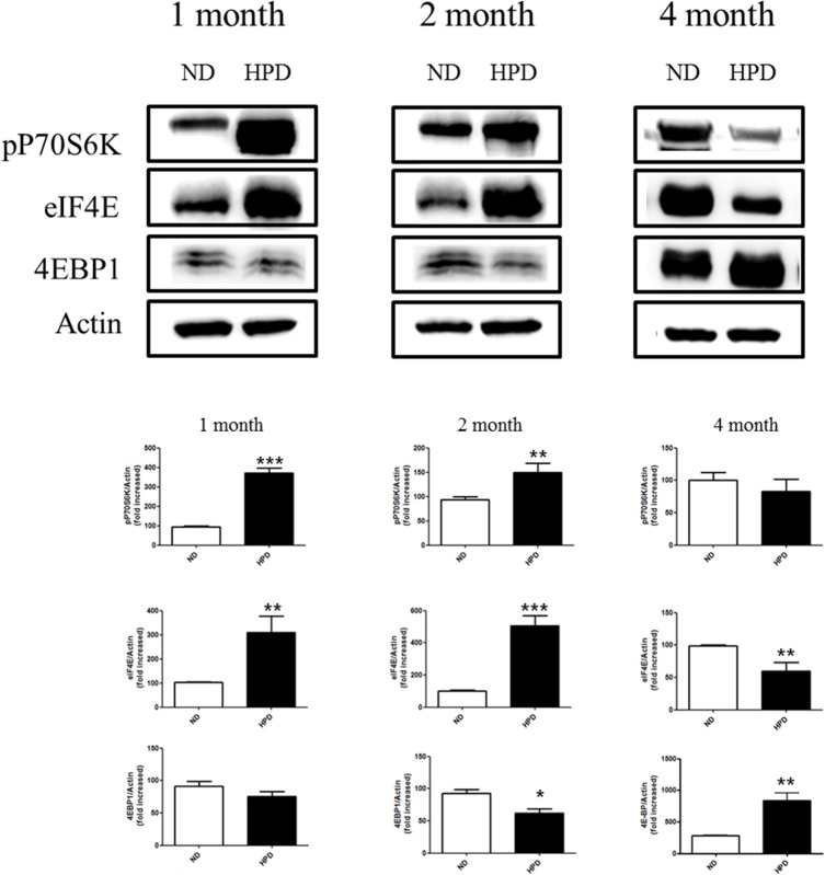
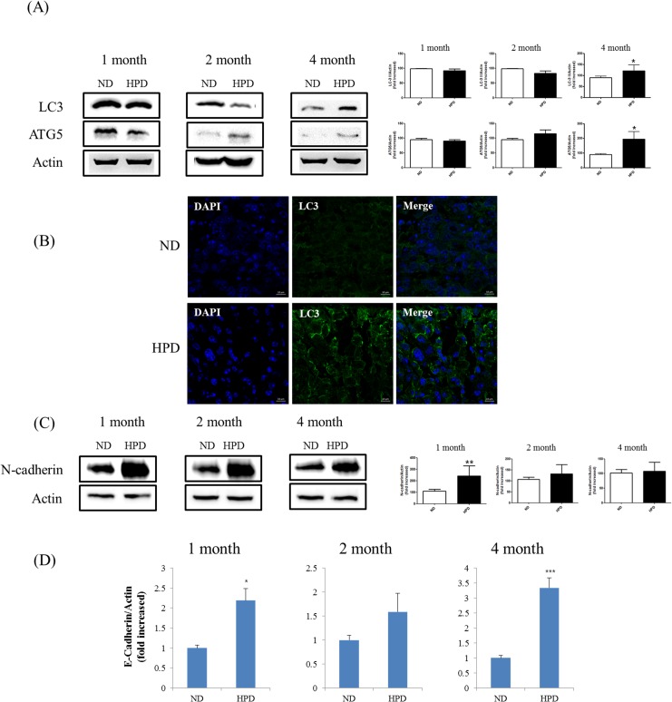
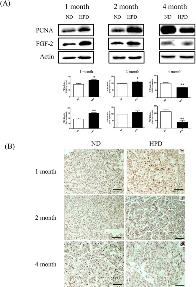
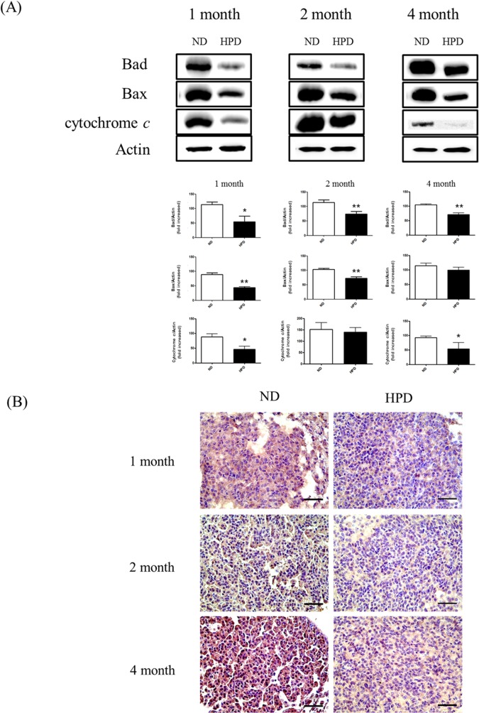
Inorganic phosphate (Pi) is required by all living organisms for the development of organs such as bone, muscle, brain, and lungs, regulating the expression of several critical genes as well as signal transduction. However, little is known about the effects of prolonged dietary Pi consumption on lung cancer progression. This study investigated the effects of a high-phosphate diet (HPD) in a mouse model of adenocarcinoma. K-rasLA1 mice were fed a normal diet (0.3% Pi) or an HPD (1% Pi) for 1, 2, or 4 months. Mice were then sacrificed and subjected to inductively coupled plasma mass/optical emission spectrometry and laser ablation inductively coupled plasma mass-spectrometry analyses, western blot analysis, histopathological, immunohistochemical, and immunocytochemical analyses to evaluate tumor formation and progression (including cell proliferation, angiogenesis, and apoptosis), changes in ion levels and metabolism, autophagy, epithelial-to-mesenchymal transition, and protein translation in the lungs. An HPD accelerated tumorigenesis, as evidenced by increased adenoma and adenocarcinoma rates as well as tumor size. However, after 4 months of the HPD, cell proliferation was arrested, and marked increases in liver and lung ion levels and in energy production via the tricarboxylic acid cycle in the liver were observed, which were accompanied by increased autophagy and decreased angiogenesis and apoptosis. These results indicate that an HPD initially promotes but later inhibits lung cancer progression because of metabolic adaptation leading to tumor cell quiescence. Moreover, the results suggest that carefully regulated Pi consumption are effective in lung cancer prevention.


Conflict of interest statement

Competing Interests: The authors have declared that no competing interests exist.

Figures

Fig 1
Fig 1. Tumor incidence in the lungs of K-rasLA1 mice provided with an ND or HPD.
Mice (n = 6 per group) were fed an ND (0.3% Pi) or HPD (1% Pi) for 1, 2, or 4 months. (A) Adenocarcinoma and adenoma in the lung tissue. (B) Total number of tumors, number of tumors with diameter >1.5 mm, and tumor volume in ND- and HPD-fed mice. The results are mean ± standard deviation (SD) of six independent measurements. Error bar represent SD. *p < 0.05; **p < 0.01; ***p < 0.001. (C) Tumors on the lungs of ND- and HPD-fed mice, as visualized by H&E staining. Scale bar: 100 μm.
Fig 2
Fig 2. Changes in ion levels in the liver and lungs of K-rasLA1 mice provided with an ND or HPD for 4 months.
LA-ICP-MS analyses represent mean values from three trials of solution ICP-MS/ICP-OES data (n = 6 per group). The vertical bar in graded color from blue to red represents the ion level from low to high, respectively.
Fig 3
Fig 3. Western blot analysis of cellular metabolism-related proteins.
Protein expression of p-acetyl CoA, acetyl CoA, SDHA (complex II), cytochrome c (complex III), COX IV (complex IV), and Rieske (Fe-S complex) relative to glyceraldehyde-3-phosphate dehydrogenase (GAPDH) in liver tissue homogenates of K-rasLA1 mice fed an ND or HPD for 1, 2, or 4 months (n = 6 per group), as determined by western blotting and quantified by densitometry. The results are mean ± SD. Error bar represent SD. *p < 0.05; **p < 0.01.
Fig 4
Fig 4. Western blot analysis of EMT- and autophagy-related proteins and qRT-PCR analysis of epithelial marker.
(A) Protein expression of the autophagy markers LC3 and ATG5 in lung tissue homogenates as determined by western blotting, using actin as a loading control. (B) LC3 expression (green) in lung tissue sections was assessed by immunocytochemistry. The highest immunoreactivity was observed in the 4-month HPD group relative to the corresponding control (ND) group. Scale bar: 10 μm. (C) Protein expression of the EMT marker N-cadherin in lung tissue homogenates of K-rasLA1 mice fed an ND or HPD for 1, 2, or 4 months (n = 6 per group), as determined by western blotting, using actin as a loading control. The results are mean ± standard deviation SD. Error bar represent SD. *p < 0.05; **p < 0.01; ***p < 0.001. (D) qRT-PCR analysis of epithelial markers, E-Cadherin. The results are mean ± standard deviation SD. Error bar represent SD. *p < 0.05; ***p < 0.001.
Fig 5
Fig 5. Analysis of protein translation in the lungs of K-rasLA1 mice provided with an ND or HPD.
The expression of the translation-related proteins p70S6K, eIF4E, p-4E-BP-1/2, and 4E-BP-1/2 in lung tissue homogenates of K-rasLA1 mice fed an ND or HPD for 1, 2, or 4 months was determined by western blotting and quantified by densitometry (n = 6 per group) relative to the expression level of actin. The results are mean ± standard deviation SD. Error bar represent SD. *p < 0.05; **p < 0.01; ***p < 0.001.
Fig 6
Fig 6. Analysis of proliferation and angiogenesis.
Proliferation and angiogenesis were evaluated in the lungs of K-rasLA1 mice fed an ND or HPD for 1, 2, or 4 months (n = 6 per group). (A) Expression of proteins associated with proliferation (PCNA) and angiogenesis (FGF-2) in lung tissue homogenates was evaluated by western blotting, using actin as a loading control. (B) PCNA-expressing cells in the tumor region (upper right corner of each panel) were visualized by immunohistochemistry. Scale bar: 20 μm. The results are mean ± standard deviation SD. Error bar represent SD. *p < 0.05; **p < 0.01.
Fig 7
Fig 7. Apoptosis in the lungs of K-rasLA1 mice provided with an ND or HPD.
Apoptosis in the lungs was analyzed in K-rasLA1 mice fed an ND or HPD for 1, 2, or 4 months (n = 6 per group). (A) The expression of the mitochondrial apoptosis-related proteins Bad, Bax, and cytochrome c was evaluated by western blotting of lung tissue homogenates, with actin used as a loading control. (B) Immunohistochemical analysis of caspase-3 expression in the tumor region. Scale bar: 20 μm. The results are mean ± standard deviation SD. Error bar represent SD. *p < 0.05; **p < 0.01.

References

    1. Takeda E, Yamamoto H, Nashiki K, Sato T, Arai H, Taketani Y. Inorganic phosphate homeostasis and the role of dietary phosphorus. J Cell Mol Med. 2004;8: 191–200. - PMC - PubMed
    1. Lampila LE. Applications and functions of food-grade phosphates. Ann N Y Acad Sci. 2013;1301: 37–44. 10.1111/nyas.12230 - DOI - PubMed
    1. Calvo MS. Dietary phosphorus, calcium metabolism and bone. J Nutr. 1993;123: 1627–1633. - PubMed
    1. Wulaningsih W, Michaelsson K, Garmo H, Hammar N, Jungner I, Walldius G, et al. Inorganic phosphate and the risk of cancer in the Swedish AMORIS study. BMC Cancer. 2013;13: 257 10.1186/1471-2407-13-257 - DOI - PMC - PubMed
    1. Chang SH, Yu KN, Lee YS, An GH, Beck GR Jr., Colburn NH, et al. Elevated inorganic phosphate stimulates Akt-ERK1/2-Mnk1 signaling in human lung cells. Am J Respir Cell Mol Biol. 2006;35: 528–539. - PMC - PubMed

Publication types

MeSH terms

LinkOut - more resources