

Quantitative theory for the longitudinal relaxation time of blood water
- PMID: 26285144
- PMCID: PMC4758918
- DOI: 10.1002/mrm.25875
Quantitative theory for the longitudinal relaxation time of blood water
Abstract
Purpose: To propose and evaluate a model for the blood water T1 that takes into account the effects of hematocrit fraction, oxygenation fraction, erythrocyte hemoglobin concentration, methemoglobin fraction, and plasma albumin concentration.
Methods: Whole blood and lysed blood T1 data were acquired at magnetic fields of 3 Tesla (T), 7T, 9.4T, and 11.7T using inversion-recovery measurements and a home-built blood circulation system for maintaining physiological conditions. A quantitative model was derived based on multivariable fitting of this data.
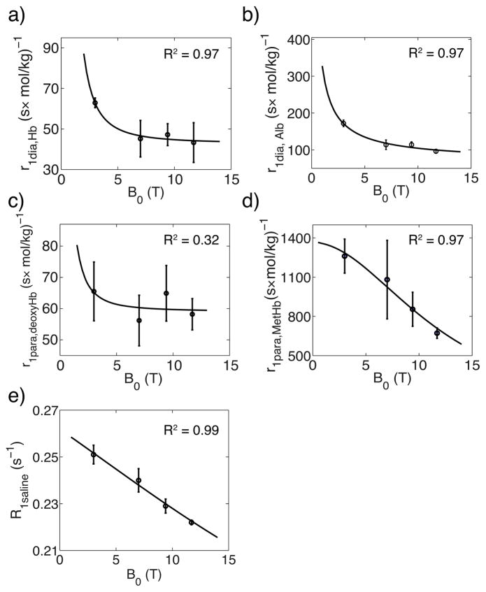
Results: Fitting of the model to the data allowed determination of the different parameters describing the blood water T1 such as those for the diamagnetic and paramagnetic effects of albumin and hemoglobin, and the contribution of methemoglobin. The model correctly predicts blood T1 at multiple fields, as verified by comparison with existing literature.
Conclusion: The model provides physical and physiological parameters describing the effects of hematocrit fraction, oxygenation, hemoglobin concentration, methemoglobin fraction, and albumin concentration on blood water T1 . It can be used to predict blood T1 at multiple fields. Magn Reson Med 76:270-281, 2016. © 2015 Wiley Periodicals, Inc.
Keywords: Hct; T1 model; albumin; hematocrit fraction; hemoglobin concentration; in vitro blood; longitudinal relaxation; methemoglobin fraction; oxygenation; relaxivity.
© 2015 Wiley Periodicals, Inc.
Figures
Similar articles
-
Quantitative theory for the transverse relaxation time of blood water.NMR Biomed. 2020 May;33(5):e4207. doi: 10.1002/nbm.4207. Epub 2020 Feb 5. NMR Biomed. 2020. PMID: 32022362 Free PMC article.
-
Transverse water relaxation in whole blood and erythrocytes at 3T, 7T, 9.4T, 11.7T and 16.4T; determination of intracellular hemoglobin and extracellular albumin relaxivities.Magn Reson Imaging. 2017 May;38:234-249. doi: 10.1016/j.mri.2016.12.012. Epub 2016 Dec 16. Magn Reson Imaging. 2017. PMID: 27993533 Free PMC article.
-
Hematocrit and oxygenation dependence of blood (1)H(2)O T(1) at 7 Tesla.Magn Reson Med. 2013 Oct;70(4):1153-9. doi: 10.1002/mrm.24547. Epub 2012 Nov 20. Magn Reson Med. 2013. PMID: 23169066 Free PMC article.
-
My starting point: the discovery of an NMR method for measuring blood oxygenation using the transverse relaxation time of blood water.Neuroimage. 2012 Aug 15;62(2):589-93. doi: 10.1016/j.neuroimage.2011.09.070. Epub 2011 Oct 6. Neuroimage. 2012. PMID: 22001265 Review.
-
[The involvement of nitric oxide in formation of hemoglobin oxygen-binding properties].Usp Fiziol Nauk. 2003 Apr-Jun;34(2):33-45. Usp Fiziol Nauk. 2003. PMID: 12754789 Review. Russian.
Cited by
-
Quantitative theory for the transverse relaxation time of blood water.NMR Biomed. 2020 May;33(5):e4207. doi: 10.1002/nbm.4207. Epub 2020 Feb 5. NMR Biomed. 2020. PMID: 32022362 Free PMC article.
-
Diffusion-weighted Imaging of the Abdomen during a Single Breath-hold Using Simultaneous-multislice Echo-planar Imaging.Magn Reson Med Sci. 2023 Apr 1;22(2):253-262. doi: 10.2463/mrms.mp.2021-0087. Epub 2021 Nov 2. Magn Reson Med Sci. 2023. PMID: 34732598 Free PMC article.
-
Is a timely assessment of the hematocrit necessary for cardiovascular magnetic resonance-derived extracellular volume measurements?J Cardiovasc Magn Reson. 2020 Nov 30;22(1):77. doi: 10.1186/s12968-020-00689-x. J Cardiovasc Magn Reson. 2020. PMID: 33250055 Free PMC article.
-
CMR-based blood oximetry via multi-parametric estimation using multiple T2 measurements.J Cardiovasc Magn Reson. 2017 Nov 9;19(1):88. doi: 10.1186/s12968-017-0403-1. J Cardiovasc Magn Reson. 2017. PMID: 29121971 Free PMC article.
-
Investigating the spatiotemporal characteristics of the deoxyhemoglobin-related and deoxyhemoglobin-unrelated functional hemodynamic response across cortical layers in awake marmosets.Neuroimage. 2018 Jan 1;164:121-130. doi: 10.1016/j.neuroimage.2017.03.005. Epub 2017 Mar 6. Neuroimage. 2018. PMID: 28274833 Free PMC article.
References
-
- Detre Ja, Leigh JS, Williams DS, Koretsky AP. Perfusion imaging. Magn Reson Med. 1992;23:37–45. - PubMed
-
- Edelman RR, Chien D, Kim D. Fast selective black blood MR imaging. Radiology. 1991;181:655–660. - PubMed
-
- Lu H, Golay X, Pekar JJ, Van Zijl PCM. Functional magnetic resonance imaging based on changes in vascular space occupancy. Magn Reson Med. 2003;50:263–274. - PubMed
-
- Bradley WG. MR appearance of hemorrhage in the brain. Radiology. 1993;189:15–26. - PubMed
-
- Cohen M, McGuire W, Cory D, Smith J. MR appearance of blood and blood products - an in vitro study. Am J Roentgenol. 1986;146:1293–1297. - PubMed
Publication types
MeSH terms
Substances
Grants and funding
LinkOut - more resources
Full Text Sources
Other Literature Sources
Medical
