

Ablation of Doublecortin-Like Kinase 1 in the Colonic Epithelium Exacerbates Dextran Sulfate Sodium-Induced Colitis
- PMID: 26285154
- PMCID: PMC4540568
- DOI: 10.1371/journal.pone.0134212
Ablation of Doublecortin-Like Kinase 1 in the Colonic Epithelium Exacerbates Dextran Sulfate Sodium-Induced Colitis
Abstract
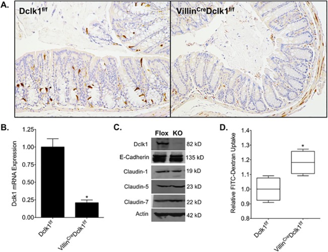
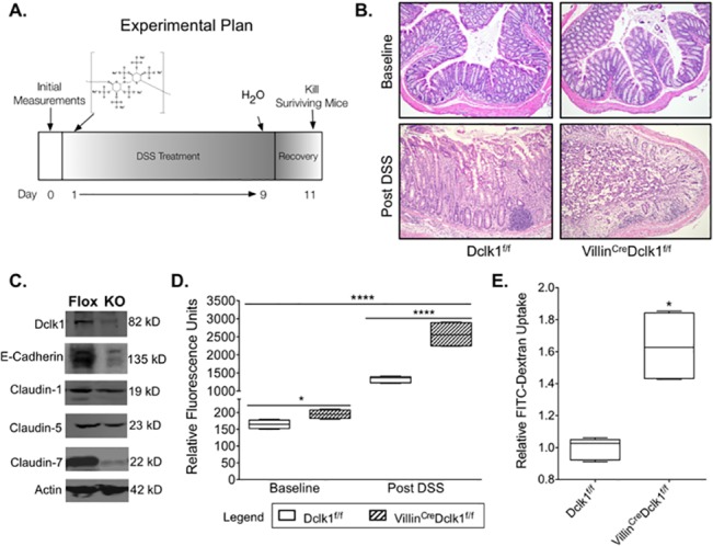
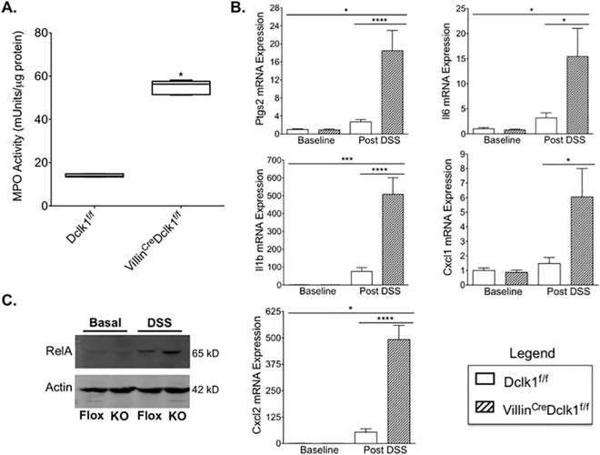
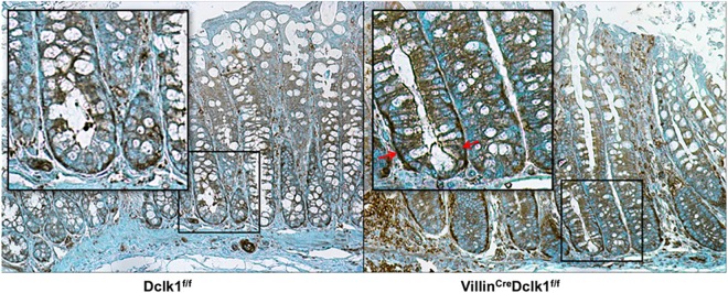
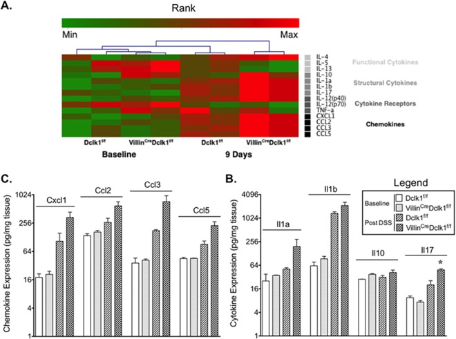
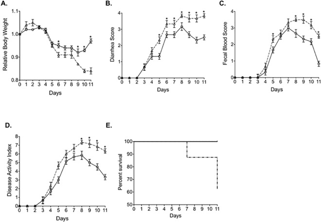
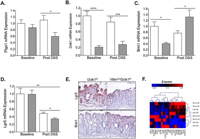
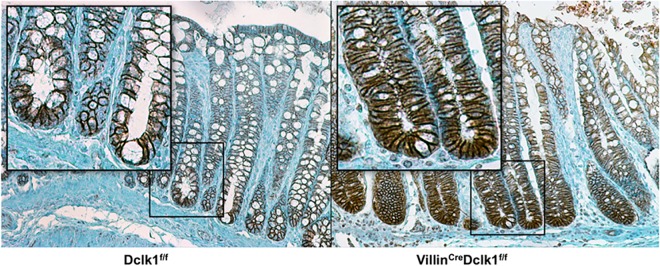
Doublecortin-like kinase 1 (Dclk1), a microtubule-associated kinase, marks the fifth lineage of intestinal epithelial cells called tuft cells that function as tumor stem cells in Apc mutant models of colon cancer. In order to determine the role of Dclk1 in dextran sulfate sodium (DSS) induced colonic inflammation both intestinal epithelial specific Dclk1 deficient (VillinCre;Dclk1f/f) and control (Dclk1f/f) mice were fed 3% DSS in drinking water for 9 days, allowed to recover for 2 days, and killed. The clinical and histological features of DSS-induced colitis were scored and immunohistochemical, gene expression, pro-inflammatory cytokines/chemokines, and immunoblotting analyses were used to examine epithelial barrier integrity, inflammation, and stem and tuft cell features. In DSS-induced colitis, VillinCre;Dclk1f/f mice demonstrated exacerbated injury including higher clinical colitis scores, increased epithelial barrier permeability, higher levels of pro-inflammatory cytokines and chemokines, decreased levels of Lgr5, and dysregulated Wnt/b-Catenin pathway genes. These results suggest that Dclk1 plays an important role in regulating colonic inflammatory response and colonic epithelial integrity.
Conflict of interest statement
Figures

Similar articles
-
Dclk1 in tuft cells promotes inflammation-driven epithelial restitution and mitigates chronic colitis.Cell Death Differ. 2019 Sep;26(9):1656-1669. doi: 10.1038/s41418-018-0237-x. Epub 2018 Nov 26. Cell Death Differ. 2019. PMID: 30478383 Free PMC article.
-
Mucosa repair mechanisms of Tong-Xie-Yao-Fang mediated by CRH-R2 in murine, dextran sulfate sodium-induced colitis.World J Gastroenterol. 2018 Apr 28;24(16):1766-1778. doi: 10.3748/wjg.v24.i16.1766. World J Gastroenterol. 2018. PMID: 29713130 Free PMC article.
-
Matrix metalloproteinase 9-induced increase in intestinal epithelial tight junction permeability contributes to the severity of experimental DSS colitis.Am J Physiol Gastrointest Liver Physiol. 2015 Dec 15;309(12):G988-97. doi: 10.1152/ajpgi.00256.2015. Epub 2015 Oct 29. Am J Physiol Gastrointest Liver Physiol. 2015. PMID: 26514773 Free PMC article.
-
Dclk1-expressing tuft cells: critical modulators of the intestinal niche?Am J Physiol Gastrointest Liver Physiol. 2017 Oct 1;313(4):G285-G299. doi: 10.1152/ajpgi.00073.2017. Epub 2017 Jul 6. Am J Physiol Gastrointest Liver Physiol. 2017. PMID: 28684459 Free PMC article. Review.
-
Advances in tuft cells, a chemosensory cell in sequential diseases of the pancreas.Biochim Biophys Acta Rev Cancer. 2023 Jul;1878(4):188911. doi: 10.1016/j.bbcan.2023.188911. Epub 2023 May 12. Biochim Biophys Acta Rev Cancer. 2023. PMID: 37182665 Review.
Cited by
-
DCLK1 and its interaction partners: An effective therapeutic target for colorectal cancer.Oncol Lett. 2021 Dec;22(6):850. doi: 10.3892/ol.2021.13111. Epub 2021 Oct 26. Oncol Lett. 2021. PMID: 34733368 Free PMC article. Review.
-
DCLK1 Expression in Colorectal Polyps Increases with the Severity of Dysplasia.In Vivo. 2018 Mar-Apr;32(2):365-371. doi: 10.21873/invivo.11247. In Vivo. 2018. PMID: 29475922 Free PMC article.
-
Type 2 immunity in intestinal homeostasis and inflammatory bowel disease.Biochem Soc Trans. 2021 Nov 1;49(5):2371-2380. doi: 10.1042/BST20210535. Biochem Soc Trans. 2021. PMID: 34581755 Free PMC article. Review.
-
Dclk1 in tuft cells promotes inflammation-driven epithelial restitution and mitigates chronic colitis.Cell Death Differ. 2019 Sep;26(9):1656-1669. doi: 10.1038/s41418-018-0237-x. Epub 2018 Nov 26. Cell Death Differ. 2019. PMID: 30478383 Free PMC article.
-
Optimized multiplex immunofluorescence single-cell analysis reveals tuft cell heterogeneity.JCI Insight. 2017 Jun 2;2(11):e93487. doi: 10.1172/jci.insight.93487. eCollection 2017 Jun 2. JCI Insight. 2017. PMID: 28570279 Free PMC article.
References
-
- Xavier RJ, Podolsky DK Unravelling the pathogenesis of inflammatory bowel disease. Nature. 2007; 448: 427–434. - PubMed
Publication types
MeSH terms
Substances
Grants and funding
LinkOut - more resources
Full Text Sources
Other Literature Sources
Molecular Biology Databases
