. 2016 Jun;87(6):650-5.
doi: 10.1136/jnnp-2015-310853. Epub 2015 Aug 18.

Management of spontaneous intracranial hypotension - Transorbital ultrasound as discriminator

Affiliations

Management of spontaneous intracranial hypotension - Transorbital ultrasound as discriminator

Jens Fichtner et al. J Neurol Neurosurg Psychiatry. 2016 Jun.

Abstract

Objective: Spontaneous intracranial hypotension (SIH) is most commonly caused by cerebrospinal fluid (CSF) leakage. Therefore, we hypothesised that patients with orthostatic headache (OH) would show decreased optic nerve sheath diameter (ONSD) during changes from supine to upright position.

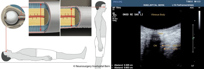
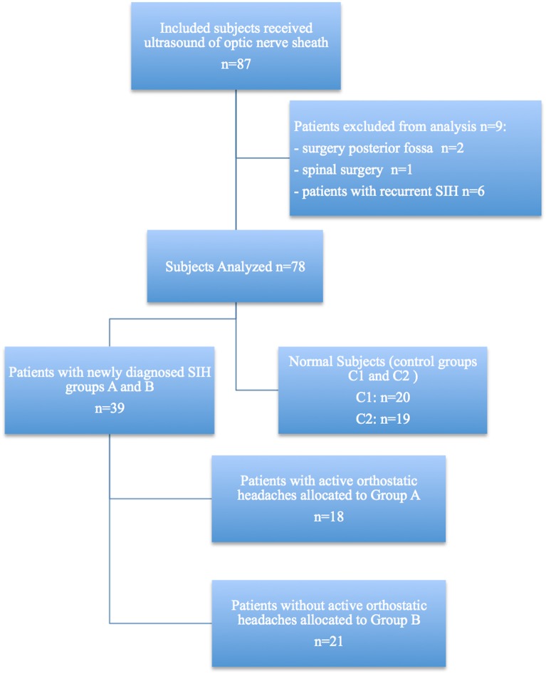
Methods: Transorbital B-mode ultrasound was performed employing a high-frequency transducer for ONSD measurements in the supine and upright positions. Absolute values and changes of ONSD from supine to upright were assessed. Ultrasound was performed in 39 SIH patients, 18 with OH and 21 without OH, and in 39 age-matched control subjects. The control group comprised 20 patients admitted for back surgery without headache or any orthostatic symptoms, and 19 healthy controls.

Results: In supine position, mean ONSD (±SD) was similar in patients with (5.38±0.91 mm) or without OH (5.48±0.89 mm; p=0.921). However, in upright position, mean ONSD was different between patients with (4.84±0.99 mm) and without OH (5.53±0.99 mm; p=0.044). Furthermore, the change in ONSD from supine to upright position was significantly greater in SIH patients with OH (-0.53±0.34 mm) than in SIH patients without OH (0.05±0.41 mm; p≤0.001) or in control subjects (0.01±0.38 mm; p≤0.001; area under the curve: 0.874 in receiver operating characteristics analysis).

Conclusions: Symptomatic patients with SIH showed a significant decrease of ONSD, as assessed by ultrasound, when changing from the supine to the upright position. Ultrasound assessment of the ONSD in two positions may be a novel, non-invasive tool for the diagnosis and follow-up of SIH and for elucidating the pathophysiology of SIH.


Figures

Figure 1
Figure 1
Left: Illustration of change in diameter of the ONS in a patient with spontaneous intracranial hypotension. Right: optic nerve sheath diameter (ONSD) image from transorbital ultrasound. Illustration of the changes in the diameter of the ONS in a symptomatic patient that occur with the change from supine to upright body position. The red arrow indicates the subarachnoid space around the optic nerve measured by ultrasound. The upper left picture shows the diameter of the ONS in a symptomatic patient while supine. The upper right picture shows a decreased ONS diameter when this patient assumes upright position. Right-side image: ONSD was assessed 3 mm behind the papilla in axial and sagittal planes on both sides using a perpendicular axis. The field of view is adjusted to a depth of 40 mm. (SAS/T=subarachnoid space/trabeculae; ONS, optic nerve sheath).
Figure 2
Figure 2
Subject disposition diagram. SIH, spontaneous intracranial hypotension.
Figure 3
Figure 3
Bee swarm box-plot data with means and SDs showing the differences between groups. Bee swarm box-plot data with means and SDs illustrate the differences (Δ) in patients with orthostatic headaches (Group A, green), without (Group B, red) and patients without an spontaneous intracranial hypotension (SIH) diagnosis (C1, grey, internal control and C2, black, external control).

References

    1. Schievink WI, Meyer FB, Atkinson JL, et al. Spontaneous spinal cerebrospinal fluid leaks and intracranial hypotension. J Neurosurg 1996;84:598–605. 10.3171/jns.1996.84.4.0598 - DOI - PubMed
    1. Headache Classification Committee of the International Headache Society (IHS). The International Classification of Headache Disorders, 3rd edition (beta version). Cephalalgia 2013;33:629–808. 10.1177/0333102413485658 - DOI - PubMed
    1. Headache Classification Subcommittee of the International Headache Society. The International Classification of Headache Disorders: 2nd edition. Cephalalgia 2004;24(Suppl 1):9–160. 10.1111/j.1468-2982.2003.00824.x - DOI - PubMed
    1. Ertl M, Barinka F, Torka E, et al. Ocular color-coded sonography—a promising tool for neurologists and intensive care physicians. Ultraschall Med 2014;5:422–31. 10.1055/s-0034-1366113 - DOI - PubMed
    1. Hansen HC, Helmke K. Validation of the optic nerve sheath response to changing cerebrospinal fluid pressure: ultrasound findings during intrathecal infusion tests. J Neurosurg 1997;87:34–40. 10.3171/jns.1997.87.1.0034 - DOI - PubMed