. 2015 Oct;4(10):1234-45.
doi: 10.5966/sctm.2015-0049. Epub 2015 Aug 18.

Targeted Disruption of the β2-Microglobulin Gene Minimizes the Immunogenicity of Human Embryonic Stem Cells

Affiliations

Targeted Disruption of the β2-Microglobulin Gene Minimizes the Immunogenicity of Human Embryonic Stem Cells

Dachun Wang et al. Stem Cells Transl Med. 2015 Oct.

Abstract

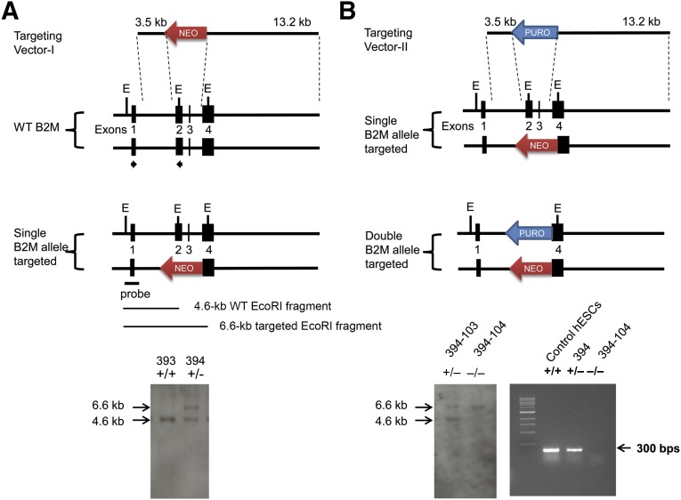
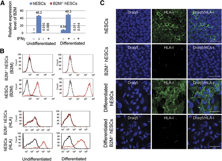
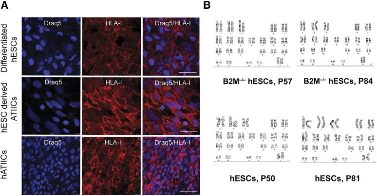
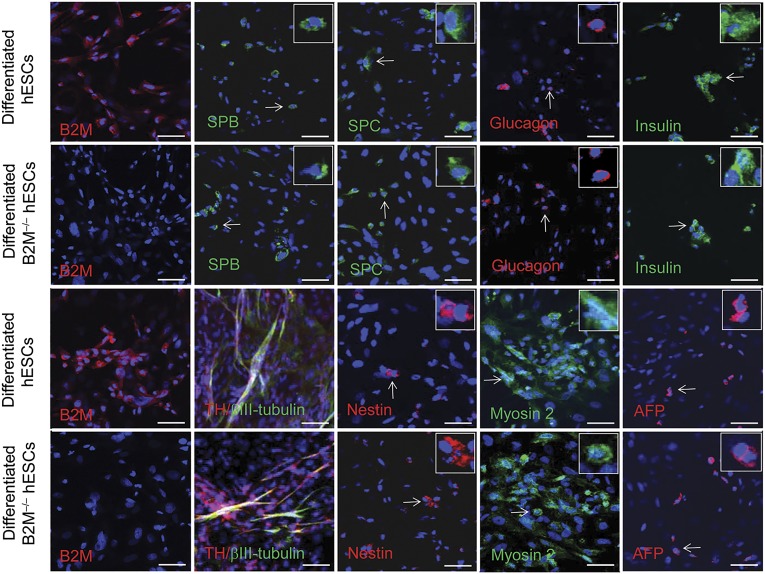
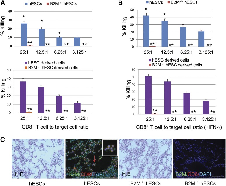
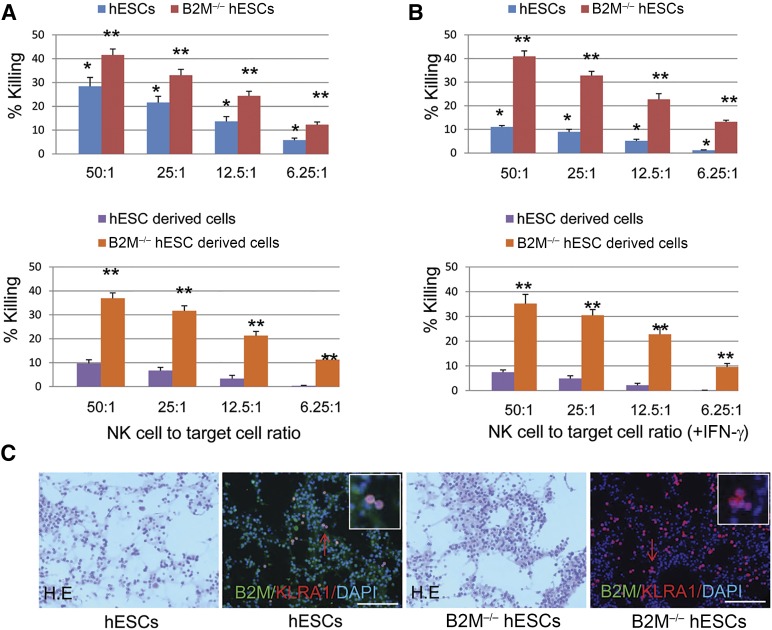
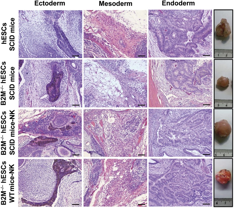
Human embryonic stem cells (hESCs) are a promising source of cells for tissue regeneration, yet histoincompatibility remains a major challenge to their clinical application. Because the human leukocyte antigen class I (HLA-I) molecules are the primary mediators of immune rejection, we hypothesized that cells derived from a hESC line lacking HLA-I expression could be transplanted without evoking a robust immune response from allogeneic recipients. In the present study, we used the replacement targeting strategy to delete exons 2 and 3 of β2-microglobulin on both gene alleles in hESCs. Because β2-microglobulin serves as the HLA-I light chain, disruption of the β2-microglobulin gene led to complete HLA-I deficiency on the cell surface of hESCs and their derivatives. Therefore, these cells were resistant to CD8+ T-cell-mediated destruction. Although interferon-γ (IFN-γ) treatment significantly induced β2-microglobulin expression, promoting CD8+ T cell-mediated killing of control hESCs and their derivatives, CD8+ T-cell-mediated cytotoxicity was barely observed with β2-microglobulin-null hESCs and their derivatives treated with IFN-γ. This genetic manipulation to disrupt HLA-I expression did not affect the self-renewal capacity, genomic stability, or pluripotency of hESCs. Despite being relatively sensitive to natural killer (NK) cell-mediated killing due to the lack of HLA-I expression, when transplanted into NK cell-depleted immunocompetent mice, β2-microglobulin-null hESCs developed into tumors resembling those derived from control hESCs in severe combined immunodeficiency mice. These results demonstrate that β2-microglobulin-null hESCs significantly reduce immunogenicity to CD8+ T cells and might provide a renewable source of cells for tissue regeneration without the need for HLA matching in the future.

Significance: This study reports the generation of a novel β2-microglobulin (B2M)-/- human embryonic stem cell (hESC) line. Differentiated mature cells from this line do not express cell surface human leukocyte antigen molecules even after interferon-γ stimulation and are resistant to alloreactive CD8+ T cells. Moreover, this B2M-/- hESC line contains no off-target integration or cleavage events, is devoid of stable B2M mRNA, exhibits a normal karyotype, and retains its self-renewal capacity, genomic stability, and pluripotency. Although B2M-/- hESC-derived cells are more susceptible to natural killer (NK) cells, murine transplantation studies have indicated that they are, overall, much less immunogenic than normal hESCs. Thus, these data show for the first time that, in vivo, the advantages provided by B2M-/- hESC-derived cells in avoiding CD8+ T-cell killing appear significantly greater than any disadvantage caused by increased susceptibility to NK cells.

Keywords: Differentiation and characterization; Human leukocyte antigen class I; Immunogenicity of human embryonic stem cells; β2-Microglobulin gene targeting strategy.


Figures

Figure 1.
Figure 1.
Targeting strategy and identification of B2M-targeted human embryonic stem cells (hESCs). The B2M-targeting vectors harbor a NEOR (targeting vector I) or PUROR gene (targeting vector II), each flanked by a 3.5-kb left arm homologous to intron 1 of the B2M gene and a 13.2-kb right arm identical to the region downstream of exon 3, including exon 4 of the B2M gene. The probe containing exon 1 sequences is upstream of the targeted region and identifies a 4.6-kb WT EcoRI B2M fragment and a 6.6-kb targeted EcoRI B2M fragment. The arrows indicate the locations of B2M forward primer I (5′-GCC TTA GCT GTG CTC GCG CTA C-3′) and reverse primer I (5′-GTC ACA TGG TTC ACA CGG CAG GCA TAC TC-3′) used for screening of B2M-targeted hESC clones. Southern hybridization identified only a 4.6-kb WT EcoRI B2M fragment in hESC-393 ([A], bottom); a 4.6-kb WT EcoRI B2M fragment and a 6.6-kb targeted EcoRI B2M fragment were detected in hESC-394, indicating that 1 of the B2M alleles had been targeted. Southern blot analysis of hESC clones from the targeting vector II transfection showed a 4.6-kb WT EcoRI B2M fragment and a 6.6-kb targeted EcoRI B2M fragment in hESC-394-103 (B2M+/− hESCs) but only a 6.6-kb targeted EcoRI B2M fragment in hESC-394-104 (B2M−/− hESCs), demonstrating that both B2M gene alleles had been disrupted in hESC-394-104 but not in hESC-394-103 ([B], bottom left). Reverse transcription-polymerase chain reaction analysis of B2M expression in the control hESCs, hESC-394 and hESC-394-104, demonstrated no B2M mRNA detected in the hESC-394-104 ([B], bottom right). Abbreviations: B2M, β2-microglobulin; bps, base pairs; E, EcoRI; WT, wild type.
Figure 2.
Figure 2.
Expression of B2M and HLA-I in B2M-null hESCs. (A): Quantitative reverse transcription-polymerase chain reaction analysis of B2M expression levels in undifferentiated and differentiated B2M−/− hESCs (hESC-394-104), with and without IFN-γ treatment, using the following primer pairs and probes: B2M forward primer-II (5′-GAG TGC TGT CTC CAT GTT TGA TG-3′), B2M reverse primer-II (5′-AAG TTG CCA GCC CTC CTA GA-3′), and B2M probe (5′-6-FAM-CTA CCT GTG GAG CAA CCT GCT CAG A-TAMRA-3′); and 18S forward primer (5′-TAA CGA ACG AGA CTCTGG CAT-3′), 18S reverse primer (5′-CGG ACA TCT AAG GGC ATC ACA G-3′), and 18S probe (5′-FAM-TGG CTG AAC GCC ACT TGT CCC TCT AA-TAMRA-3′). The expression levels were normalized to endogenous 18S rRNA control. Bar graphs depict B2M mRNA expression levels relative to hESC control (n = 5). (B): Flow cytometry was performed to analyze surface B2M or HLA-I protein expression in the hESCs and B2M−/− hESCs (hESC-394-104) before and after differentiation using the mouse anti-B2M antibody (top panel, red) or mouse anti-human MHC-I antibody (bottom panel, red) with mouse IgG as the isotype control (black). (C): Immunofluorescent staining demonstrated negative HLA-I expression on the surface of undifferentiated and differentiated B2M−/− hESCs (hESC-394-104), with hESCs serving as the control. Scale bars = 50 μm (human major histocompatibility complex I antibody: 1:50 diluted). Abbreviations: B2M, β2-microglobulin; hESCs, human embryonic stem cells; HLA, human leukocyte antigen; IFN-γ, interferon-γ.
Figure 3.
Figure 3.
HLA-I expression in hESC-derived ATIICs and cytogenetic analysis B2M-null hESCs. (A): Immunofluorescent staining for surface HLA-I expression on differentiated hESCs, hESC-derived ATIICs, and hATIICs using 1:150 diluted human major histocompatibility complex I antibody. Scale bars = 50 μm. (B): Cytogenetic evaluation of B2M−/− hESCs (hESC-394-104) with hESC control. Twenty metaphase cells harvested from each cell type were analyzed at different passages. Abbreviations: ATIICs, alveolar epithelial type II cells; B2M, β2-microglobulin; hATIICs, human primary ATIICs; hESCs, human embryonic stem cells; HLA, human leukocyte antigen.
Figure 4.
Figure 4.
In vitro differentiation of B2M-null hESCs. Immunofluorescent staining of differentiated cultures of B2M-null hESCs was performed to examine their potential to differentiate into various cell lineages from three germ layers with hESC control. The differentiated B2M-null hESCs were B2M negative compared with the differentiated control hESCs (first column images). The SPB- or SPC-expressing distal lung epithelial cell types (green), glucagon-positive pancreatic α cells (red), insulin-producing pancreatic β cells (green), and AFP-positive hepatocyte progenitor cells (red) derived from endoderm were identified in the cultures (the right four images in the top two rows and the fifth images in the bottom two rows). Mesoderm-derived muscle cells showing positive myosin-2 staining were demonstrated in the differentiated cultures (fourth images in the bottom two rows). The ability of B2M-null hESCs to differentiate into ectoderm-derived cells was demonstrated by derived nestin, TH, and/or βIII-tubulin-positive nerve cells in the cultures (second and third images in the bottom two rows). A magnified view of a representative B2M-null hESC-derived cell indicated by a white arrow is inset in each corresponding image. Scale bars = 50 μm. Abbreviations: AFP, α1-fetoprotein; B2M, β2-microglobulin; hESCs, human embryonic stem cells; SPB, surfactant protein B; SPC, surfactant protein C; TH, tyrosine hydroxylase.
Figure 5.
Figure 5.
Resistance of B2M-null hESCs and their derivatives to alloreactive CD8+ T cell-mediated killing in vitro. Histogram representation of the cytotoxic effects of CD8+ T cells on undifferentiated and differentiated B2M−/− hESCs (A) and on IFN-γ-treated B2M−/− hESCs (B) before and after differentiation with hESC control. The various ratios of CD8+ T cell to target cell are indicated (n = 5; ∗, p < .01 vs. differentiated hESCs with or without IFN-γ treatment; ∗∗, p < .0001 vs. undifferentiated or differentiated hESCs). (C): 4 × 10⁶ hESCs or B2M−/− hESCs were mixed with 200 ml of growth factor-reduced Matrigel and then injected subcutaneously into 10-week-old immunocompetent mice. Implants were harvested after 48 hours and then fixed in 4% formalin, sectioned, and stained with mouse anti-B2M (1:100 dilution; Santa Cruz Biotechnology) and rabbit anti-CD8 antibody (1:100 dilution; Bioss Inc., Woburn, MA, http://www.biossusa.com) or with H&E. Representative immunofluorescence detection of numerous CD8+ T cells (red) infiltrating into B2M-positive (green) Matrigel-hESC implants shown (second image), but rare CD8+ T cells were present in the B2M-negative Matrigel-B2M−/− hESC implants (fourth image). A magnified view of representative CD8+ T cells indicated by red arrow was inserted in each corresponding image. Scale bars = 100 μm. Abbreviations: B2M, β2-microglobulin; DAPI, 4′,6-diamidino-2-phenylindole; hESC, human embryonic stem cell; IFN-γ, interferon-γ.
Figure 6.
Figure 6.
Susceptibility of B2M-null hESCs to NK cell-mediated killing. Bar graph representation of the cytotoxic effects of NK cells on undifferentiated and differentiated B2M−/− hESCs (A) and IFN-γ-treated B2M−/− hESCs before and after differentiation (B) with hESC control. The various ratios of NK cell to target cell are indicated (n = 5; ∗, p < .01 vs. differentiated hESCs treated with or without IFN-γ; ∗∗, p < .01 vs. undifferentiated or differentiated hESCs). (C): 4 × 10⁶ hESCs or B2M−/− hESCs were mixed with 200 ml of growth factor-reduced Matrigel and then injected subcutaneously into 10-week-old immunocompetent mice. Implants were harvested after 48 hours and then fixed in 4% formalin, sectioned, and stained with mouse anti-B2M and rabbit anti-mouse KLRA1 antibody (1:100 dilution; Abcam) or with H&E. Representative immunofluorescence detection of KLRA1-positive NK cells (red) infiltrating into the B2M-positive (green) Matrigel-hESC implants or B2M-negative Matrigel-B2M−/− hESC implants shown. A magnified view of representative KLRA1+ cells indicated by red arrow was inserted in each corresponding image. Scale bars = 100 μm. Abbreviations: B2M, β2-microglobulin; DAPI, 4′,6-diamidino-2-phenylindole; hESC, human embryonic stem cell; IFN-γ, interferon-γ; NK, natural killer.
Figure 7.
Figure 7.
Immunogenicity and pluripotency of B2M-null hESCs in immunocompetent mice. The ability of B2M−/− hESCs (hESC-394-104) to evade the immune response was examined by teratoma formation assay in SCID and immunocompetent mice with and without NK cell depletion using hESCs as control. As hESCs, B2M−/− hESCs developed into tumors in SCID mice, but the teratoma sizes were significantly smaller than those derived from hESCs due to NK cell-mediated killing (second row, fourth image from left). However, the teratomas derived from B2M−/− hESCs in SCID and immunocompetent mice resembled those derived from control hESCs in SCID mice provided that NK cells were depleted before transplantation (fourth images in bottom two rows). H&E staining of representative teratoma sections showed various tissues derived from hESCs and B2M−/− hESCs. Scale bars = 200 μm. Abbreviations: B2M, β2-microglobulin; hESC, human embryonic stem cell; IFN-γ, interferon-γ; NK, natural killer; SCID, severe combined immunodeficiency; WT, wild type.

References

    1. Thomson JA, Itskovitz-Eldor J, Shapiro SS, et al. Embryonic stem cell lines derived from human blastocysts. Science. 1998;282:1145–1147. - PubMed
    1. Reubinoff BE, Pera MF, Fong CY, et al. Embryonic stem cell lines from human blastocysts: Somatic differentiation in vitro. Nat Biotechnol. 2000;18:399–404. - PubMed
    1. Mummery C, Ward-van Oostwaard D, Doevendans P, et al. Differentiation of human embryonic stem cells to cardiomyocytes: Role of coculture with visceral endoderm-like cells. Circulation. 2003;107:2733–2740. - PubMed
    1. Johnson MA, Weick JP, Pearce RA, et al. Functional neural development from human embryonic stem cells: Accelerated synaptic activity via astrocyte coculture. J Neurosci. 2007;27:3069–3077. - PMC - PubMed
    1. Wang D, Haviland DL, Burns AR, et al. A pure population of lung alveolar epithelial type II cells derived from human embryonic stem cells. Proc Natl Acad Sci USA. 2007;104:4449–4454. - PMC - PubMed

Publication types

MeSH terms