

Tissue-engineered tubular substitutions for urinary diversion in a rabbit model
- PMID: 26286106
- PMCID: PMC4935384
- DOI: 10.1177/1535370215600101
Tissue-engineered tubular substitutions for urinary diversion in a rabbit model
Abstract
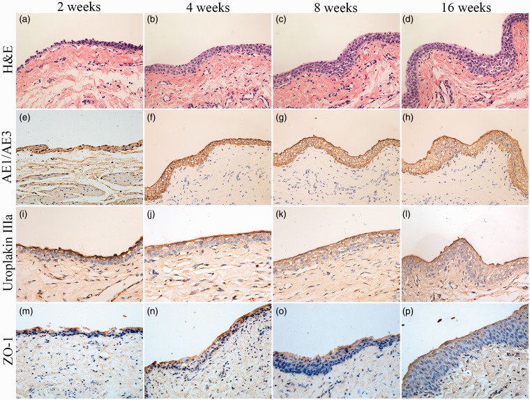
Clinically, autologous gastrointestinal segments are traditionally used for urinary diversion. However, this procedure often causes many serious complications. Tissue engineering may provide an alternative treatment method in urinary diversion. This research aims to produce tissue-engineered tubular substitutions by using homologous adipose-derived stem cells, smooth muscle cells, and bladder acellular matrix in developing urinary diversion in a rabbit model. Adipose-derived stem cells and smooth muscle cells of rabbit were obtained and cultured in vitro. These cultured adipose-derived stem cells and smooth muscle cells were seeded onto the two sides of the bladder acellular matrix and then incubated for seven days. The cell-seeded matrix was used to build tissue-engineered tubular substitutions, which were then implanted and wrapped into the omentum in vivo for two weeks to promote angiogenesis. In the experimental group, the bladder of 20 rabbits was totally resected, and the above tissue-engineered tubular substitutions were used for urinary diversion. In the control group, bladder acellular matrix tubular substitutions with unseeded cells were implanted into the omentum and were used as urinary diversion on another five rabbits with the same process. The implants were harvested, and histological examination was conducted at 2, 4, 8, and 16 weeks after operation. Intravenous urography assessment was performed at 16 weeks postoperatively. All the rabbits were alive in the experimental group until they were sacrificed. Histological analysis of the construct displayed the presence of multilayer urothelial cells on the luminal side and organized smooth muscle tissue on the other side, and different diameters of neovascularization were clearly identified in the substitutions obtained. No leakage, stricture, or obstructions were noted with intravenous urography assessment. All the animals in the control group died within two weeks, and urine leakage, scar formation, and inflammation were detected through autopsy. This study demonstrates the feasibility of tissue-engineered tubular substitutions constructed using homologous adipose-derived stem cells, smooth muscle cells, and bladder acellular matrix for urinary diversion in a rabbit model.
Keywords: Tissue engineering; adipose-derived stem cells; bladder acellular matrix; epithelium; omentum; smooth muscle cells; urinary diversion.
© 2015 by the Society for Experimental Biology and Medicine.
Figures


Similar articles
-
Construction of ureteral grafts by seeding bone marrow mesenchymal stem cells and smooth muscle cells into bladder acellular matrix.Transplant Proc. 2013 Mar;45(2):730-4. doi: 10.1016/j.transproceed.2012.08.023. Transplant Proc. 2013. PMID: 23498814
-
Tissue-engineered tubular graft for urinary diversion after radical cystectomy in rabbits.J Surg Res. 2013 Jun 15;182(2):185-91. doi: 10.1016/j.jss.2012.10.024. Epub 2012 Nov 3. J Surg Res. 2013. PMID: 23140788
-
Tissue-engineered tubular substitutions for urinary diversion in a preclinical rabbit model.Front Med (Lausanne). 2025 Jun 26;12:1616977. doi: 10.3389/fmed.2025.1616977. eCollection 2025. Front Med (Lausanne). 2025. PMID: 40641968 Free PMC article.
-
Whyever bladder tissue engineering clinical applications still remain unusual even though many intriguing technological advances have been reached?G Chir. 2016 Jan-Feb;37(1):6-12. doi: 10.11138/gchir/2016.37.1.006. G Chir. 2016. PMID: 27142819 Free PMC article. Review.
-
Tissue-engineered urinary conduits.Curr Urol Rep. 2015 Mar;16(3):8. doi: 10.1007/s11934-015-0480-3. Curr Urol Rep. 2015. PMID: 25677229 Review.
Cited by
-
Decellularized scaffolds in regenerative medicine.Oncotarget. 2016 Sep 6;7(36):58671-58683. doi: 10.18632/oncotarget.10945. Oncotarget. 2016. PMID: 27486772 Free PMC article. Review.
-
Autologous micrografting improves regeneration of tissue-engineered urinary conduits in vivo.Sci Rep. 2024 Sep 25;14(1):22028. doi: 10.1038/s41598-024-72876-0. Sci Rep. 2024. PMID: 39322716 Free PMC article.
-
Stem cell therapy for bladder regeneration: A comprehensive systematic review.Regen Ther. 2024 Dec 21;28:191-200. doi: 10.1016/j.reth.2024.12.005. eCollection 2025 Mar. Regen Ther. 2024. PMID: 39811066 Free PMC article. Review.
-
Biofabrication and biomaterials for urinary tract reconstruction.Res Rep Urol. 2017 May 10;9:79-92. doi: 10.2147/RRU.S127209. eCollection 2017. Res Rep Urol. 2017. PMID: 28546955 Free PMC article. Review.
-
Evaluation of PC12 Cells' Proliferation, Adhesion and Migration with the Use of an Extracellular Matrix (CorMatrix) for Application in Neural Tissue Engineering.Materials (Basel). 2021 Jul 10;14(14):3858. doi: 10.3390/ma14143858. Materials (Basel). 2021. PMID: 34300779 Free PMC article.
References
-
- Biers SM, Venn SN, Greenwell TJ. The past, present and future of augmentation cystoplasty. BJU Int 2012; 109: 1280–93. - PubMed
-
- Scales CD, Jr, Wiener JS. Evaluating outcomes of enterocystoplasty in patients with spina bifida: a review of the literature. J Urol 2008; 180: 2323–9. - PubMed
-
- Husmann DA, Rathbun SR. Long-term follow up of enteric bladder augmentations: the risk for malignancy. J Pediatr Urol 2008; 4: 381–5; discussion 6. - PubMed
-
- Zhang Y, Atala A. Urothelial cell culture: stratified urothelial sheet and three-dimensional growth of urothelial structure. Methods Mol Biol 2013; 945: 383–99. - PubMed
-
- Horst M, Madduri S, Gobet R, Sulser T, Milleret V, Hall H, Atala A, Eberli D. Engineering functional bladder tissues. J Tissue Eng Regener Med 2013; 7: 515–22. - PubMed
Publication types
MeSH terms
LinkOut - more resources
Full Text Sources
Other Literature Sources
Medical