

Immune phenotypes of microglia in human neurodegenerative disease: challenges to detecting microglial polarization in human brains
- PMID: 26286145
- PMCID: PMC4543480
- DOI: 10.1186/s13195-015-0139-9
Immune phenotypes of microglia in human neurodegenerative disease: challenges to detecting microglial polarization in human brains
Abstract
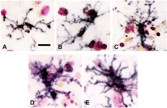
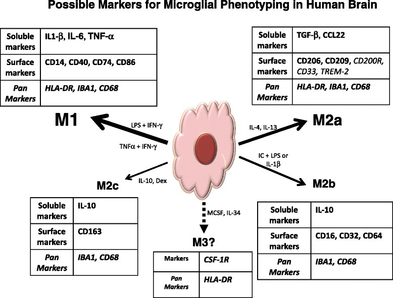
Inflammatory responses in the brain, which can be demonstrated by changes in properties of microglia, the brain-resident macrophages, are a common feature of human neurodegenerative diseases. Different monocyte/macrophage phenotypes have been defined by changes in expression of cytokines, receptors and other markers as a response to different classes of stimuli. Monocytes, macrophages and microglia can have a range of phenotypes with associated properties depending on their microenvironment. Macrophage/microglia polarization states have been defined as classical activation (M1), alternative activation (M2a), type II alternative activation (M2b) or acquired deactivation (M2c). Available markers for identifying microglial phenotypes in human brains are still limited; those available provide incomplete information on the functions or polarization states of microglia observed in tissues from diseases such as Alzheimer's disease, Parkinson's disease and multiple sclerosis. The most widely used marker to describe activated microglia in human brains, particularly diseased brains, has been HLA-DR, the major histocompatibility complex II protein. HLA-DR-positive microglia can have a wide range of activation morphologies that are affected not only by disease pathology, but also by their differentiation states and brain regions. Two other widely used markers to identify microglia in human brains are ionized calcium binding adaptor molecule-1 and CD68. Although their expression changes in diseased brains, these markers do not show specificity for different phenotypes. Over the years there have been studies with additional markers that attempt to further define microglial properties, particularly in Alzheimer's disease brains. Most studies have employed immunohistochemical techniques to identify microglia in tissue sections, but recent advances in this field have allowed gene expression profiling of microglia upon immediate isolation from brains. We will review which markers might better define different activation phenotypes of microglia in human brains and whether they fit into current microglial polarization schemes.
Figures
Similar articles
-
Patterns of Expression of Purinergic Receptor P2RY12, a Putative Marker for Non-Activated Microglia, in Aged and Alzheimer's Disease Brains.Int J Mol Sci. 2020 Jan 20;21(2):678. doi: 10.3390/ijms21020678. Int J Mol Sci. 2020. PMID: 31968618 Free PMC article.
-
TMEM119 marks a subset of microglia in the human brain.Neuropathology. 2016 Feb;36(1):39-49. doi: 10.1111/neup.12235. Epub 2015 Aug 6. Neuropathology. 2016. PMID: 26250788
-
Exploring the Significance of Microglial Phenotypes and Morphological Diversity in Neuroinflammation and Neurodegenerative Diseases: From Mechanisms to Potential Therapeutic Targets.Immunol Invest. 2024 Aug;53(6):891-946. doi: 10.1080/08820139.2024.2358446. Epub 2024 Jun 5. Immunol Invest. 2024. PMID: 38836373 Review.
-
Microglial Phenotyping in Neurodegenerative Disease Brains: Identification of Reactive Microglia with an Antibody to Variant of CD105/Endoglin.Cells. 2019 Jul 23;8(7):766. doi: 10.3390/cells8070766. Cells. 2019. PMID: 31340569 Free PMC article.
-
Expression of immunohistochemical markers on microglia in Creutzfeldt-Jakob disease and Alzheimer's disease: morphometric study and review of the literature.Folia Neuropathol. 2012;50(1):74-84. Folia Neuropathol. 2012. PMID: 22505366 Review.
Cited by
-
Neural repair function of osteopontin in stroke and stroke‑related diseases (Review).Exp Ther Med. 2024 Oct 16;28(6):459. doi: 10.3892/etm.2024.12749. eCollection 2024 Dec. Exp Ther Med. 2024. PMID: 39478739 Free PMC article. Review.
-
Human Microglia Extensively Reconstitute in Humanized-BLT Mice With Human Interleukin-34 Transgene and Support HIV-1 Brain Infection.Front Immunol. 2021 May 21;12:672415. doi: 10.3389/fimmu.2021.672415. eCollection 2021. Front Immunol. 2021. PMID: 34093573 Free PMC article.
-
A glance through the effects of CD4+ T cells, CD8+ T cells, and cytokines on Alzheimer's disease.Comput Struct Biotechnol J. 2023 Nov 8;21:5662-5675. doi: 10.1016/j.csbj.2023.10.058. eCollection 2023. Comput Struct Biotechnol J. 2023. PMID: 38053545 Free PMC article. Review.
-
Treatment with Granulocyte-Macrophage Colony-Stimulating Factor Reduces Viral Titers in the Brains of West Nile Virus-Infected Mice and Improves Survival.J Virol. 2023 Mar 30;97(3):e0180522. doi: 10.1128/jvi.01805-22. Epub 2023 Feb 21. J Virol. 2023. PMID: 36802227 Free PMC article.
-
Role of exosomes in the pathogenesis of inflammation in Parkinson's disease.Neural Regen Res. 2022 Sep;17(9):1898-1906. doi: 10.4103/1673-5374.335143. Neural Regen Res. 2022. PMID: 35142665 Free PMC article. Review.
References
-
- Akiyama H, Barger S, Barnum S, Bradt B, Bauer J, Cole GM, Cooper NR, Eikelenboom P, Emmerling M, Fiebich BL, Finch CE, Frautschy S, Griffin WS, Hampel H, Hull M, Landreth G, Lue L, Mrak R, Mackenzie IR, McGeer PL, O'Banion MK, Pachter J, Pasinetti G, Plata-Salaman C, Rogers J, Rydel R, Shen Y, Streit W, Strohmeyer R, Tooyoma I, Van Muiswinkel FL, Veerhuis R, Walker D, Webster S, Wegrzyniak B, Wenk G, Wyss-Coray T. Inflammation and Alzheimer's disease. Neurobiol Aging. 2000;21:383–421. doi: 10.1016/S0197-4580(00)00124-X. - DOI - PMC - PubMed
-
- Heneka MT, Carson MJ, Khoury JE, Landreth GE, Brosseron F, Feinstein DL, Jacobs AH, Wyss-Coray T, Vitorica J, Ransohoff RM, Herrup K, Frautschy SA, Finsen B, Brown GC, Verkhratsky A, Yamanaka K, Koistinaho J, Latz E, Halle A, Petzold GC, Town T, Morgan D, Shinohara ML, Perry VH, Holmes C, Bazan NG, Brooks DJ, Hunot S, Joseph B, Deigendesch N, Garaschuk O, Boddeke E, Dinarello CA, Breitner JC, Cole GM, Golenbock DT, Kummer MP. Neuroinflammation in Alzheimer's disease. Lancet Neurol. 2015;14:388–405. doi: 10.1016/S1474-4422(15)70016-5. - DOI - PMC - PubMed
Publication types
MeSH terms
Substances
Grants and funding
LinkOut - more resources
Full Text Sources
Other Literature Sources
Medical
Research Materials
