. 2015 Aug 19:16:19.
doi: 10.1186/s12858-015-0048-3.

Identification of inhibitors that target dual-specificity phosphatase 5 provide new insights into the binding requirements for the two phosphate pockets

Affiliations

Identification of inhibitors that target dual-specificity phosphatase 5 provide new insights into the binding requirements for the two phosphate pockets

Terrence S Neumann et al. BMC Biochem. .

Abstract

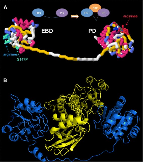
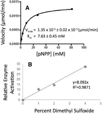
Background: Dual-specificity phosphatase-5 (DUSP5) plays a central role in vascular development and disease. We present a p-nitrophenol phosphate (pNPP) based enzymatic assay to screen for inhibitors of the phosphatase domain of DUSP5.

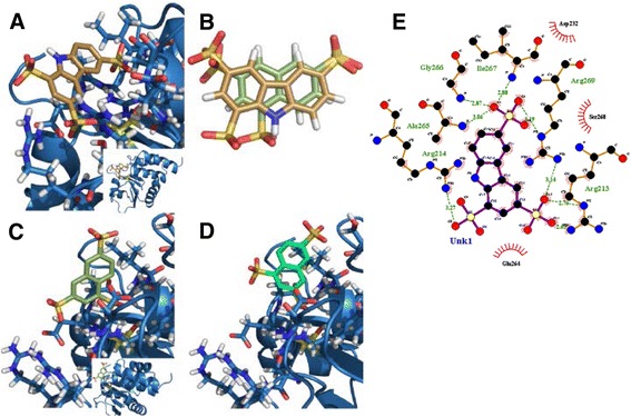
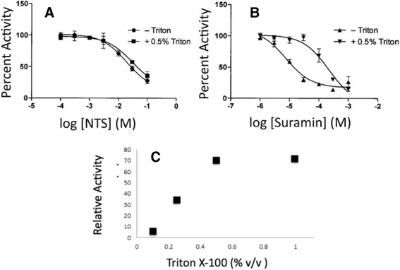
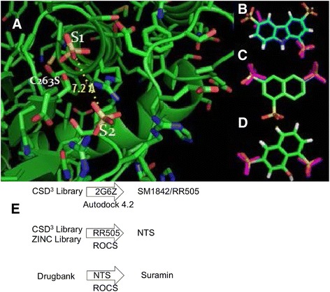
Methods: pNPP is a mimic of the phosphorylated tyrosine on the ERK2 substrate (pERK2) and binds the DUSP5 phosphatase domain with a Km of 7.6 ± 0.4 mM. Docking followed by inhibitor verification using the pNPP assay identified a series of polysulfonated aromatic inhibitors that occupy the DUSP5 active site in the region that is likely occupied by the dual-phosphorylated ERK2 substrate tripeptide (pThr-Glu-pTyr). Secondary assays were performed with full length DUSP5 with ERK2 as substrate.

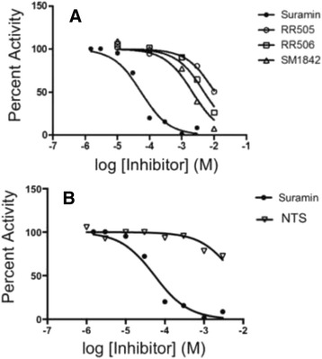
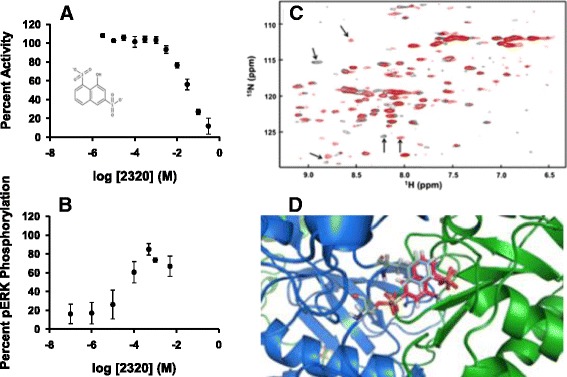
Results: The most potent inhibitor has a naphthalene trisulfonate (NTS) core. A search for similar compounds in a drug database identified suramin, a dimerized form of NTS. While suramin appears to be a potent and competitive inhibitor (25 ± 5 μM), binding to the DUSP5 phosphatase domain more tightly than the monomeric ligands of which it is comprised, it also aggregates. Further ligand-based screening, based on a pharmacophore derived from the 7 Å separation of sulfonates on inhibitors and on sulfates present in the DUSP5 crystal structure, identified a disulfonated and phenolic naphthalene inhibitor (CSD (3) _2320) with IC₅₀ of 33 μM that is similar to NTS and does not aggregate.

Conclusions: The new DUSP5 inhibitors we identify in this study typically have sulfonates 7 Å apart, likely positioning them where the two phosphates of the substrate peptide (pThr-Glu-pTyr) bind, with one inhibitor also positioning a phenolic hydroxyl where the water nucleophile may reside. Polysulfonated aromatic compounds do not commonly appear in drugs and have a tendency to aggregate. One FDA-approved polysulfonated drug, suramin, inhibits DUSP5 and also aggregates. Docking and modeling studies presented herein identify polysulfonated aromatic inhibitors that do not aggregate, and provide insights to guide future design of mimics of the dual-phosphate loops of the ERK substrates for DUSPs.


Figures

Fig. 1
Fig. 1
DUSP5 and ERK2 Models. a Model depicting the two domains of DUSP5. This model is comprised of two domains, the ERK binding domain (EBD) and phosphatase domain (PD), and illustrates the relative location of the domains and their connection via a 30 amino acid linker of unknown structure. The homology model of EBD was constructed using the solution structure (21 % identity and 35 % homology) of human MKP-3 protein (PDB:1HZM) as a template [35]. The phosphatase domain is the previously reported crystal structure (PDB:2G6Z) [16]. The 30 amino acid linker region connecting the two domains was prepared manually, and is of unknown structure. The S147P mutation present in patients with vascular anomalies is shown in green, and arginine-rich basic regions have been identified. b DUSP5 and ERK2 binding model. DUSP5 (blue) is positioned similarly in respects to panel a with the EBD to the left and PD to the right, wrapping around human ERK2 in yellow. Model was prepared as described in our previous paper [8]. The linker region may have the first 11 amino acids as helical based secondary structure predictions [–48], although this was only found to be loosely helical after molecular dynamics simulations. The ERK2 (yellow) structure (PDB:3I60) [18] is shown between the DUSP5 domains to illustrate relative shape and size complementarity; and, relative orientation of ERK2 and DUSP5 is based on the molecular dynamics simulation and associated analysis presented in our previous paper [8]
Fig. 2
Fig. 2
Docking Results. a Predicted docking pose of SM1842/RR505 (gold) in DUSP5 PD (blue), using Autodock 4.2. The inset image shows predicted binding position relative to the rest of the protein. The side chains around the bound ligand (mostly arginine guanido groups) are delineated in light turquoise and the catalytic cysteine is displayed in yellow. Three arginine residues are observed around one sulfonate group of SM1842/RR505. The calculated binding energy for this pose was -9.69 kcal/mol and had a cluster population of 10. b Optimal overlay of SM1842/RR505 (gold) and naphthalene trisulfonate (NTS, moss green), using OpenEye Scientific Software ROCS v. 3.0 [26]. c Lowest energy binding pose for NTS (moss green) in DUSP5 PD (blue), with a calculated binding energy of -8.48 kcal/mol with a cluster population of 7. d Second lowest energy binding pose for NTS (seafoam), with a calculated binding energy of -8.21 kcal/mol. e Ligplot drawing of SM1842/RR505 in the DUSP5 PD binding pocket, showing key interactions
Fig. 3
Fig. 3
Michaelis-Menten Kinetics. a Michaelis-Menten plot of DUSP5 PD(WT) initial velocity versus substrate (pNPP) concentration, monitoring production of p-nitrophenolate at 405 nm. Reaction was in 100 mM Tris-HCl (pH 7.5), 100 mM NaCl, 5 mM MgCl2 and 1 mM DTT, and was initiated with enzyme. The line represents a nonlinear least squares fit to equation 1. b Enzymatic rate as a function of DMSO concentration (% v/v), and at a fixed level of pNPP (5 mM), with other conditions as in panel (a). Relative enzyme activation represents the rate normalized to that obtained at 0 % DMSO
Fig. 4
Fig. 4
IC 50 Measurements. a DUSP5 PD(WT) initial velocity versus inhibitor concentration, and fitted to equation 3 to obtain IC50 values (Table 1). Conditions were as described for Fig. 3. (b) Same as panel (a), but comparing suramin and NTS, demonstrating the affinity increase that is obtained due to tethering the NTS fragments
Fig. 5
Fig. 5
Effect of Detergent on Suramin Inhibition. a NTS IC50 measurement in the presence and absence of 0.5 % Triton, showing no detergent-effect on inhibition of DUSP5 PD(WT) pNPP phosphatase activity. b Suramin IC50 measurement in the presence and absence of 0.5 % Triton X-100 shows a loss of some inhibitory capability in the presence of detergent. c Effect of increasing detergent levels (Triton X-100) on rate of DUSP5 PD(WT) in the presence of a fixed concentrations of inhibitor and substrate (at 1 μM suramin and 5 mM pNPP). Detergent removes some, but not all, of suramin’s inhibitory effect, showing a plateau level 30 % inhibition
Fig. 6
Fig. 6
Pharmacophore-based Identification of DUSP5 PD Inhibitors. a Crystal structure of DUSP5 PD(C263S) [16], showing the two bound sulfate ions in the two anion-binding pockets postulated to be occupied by the two phosphate groups of the ERK2 activation loop (pThr-Glu-pTyr) [–18]. The anion pocket closest to the catalytic nucleophile (Cys263) is labeled S1, and the distal anion pocket is labeled S2. The S2 anion (sulfate) is stabilized be several arginine residues, while the S1 anion may derive some helix dipole stabilization by virtue of its location at the N-terminal end of a long central helix. The sulfur to sulfur distance of 7.2 Å defines the DUSP PD pharmacophore as two anionic groups separated by ~7 Å. Overlay of the S1-S2 pharmacophore (two sulfates, shown as purple) on RR505 indicates a poor match, while (b) overlay on NTS (c) in one of two possible orientations (related by a 180° rotation) is better. d A ligand-based search using this pharmacophore identified CSD 3 _2320, which also matched the S1-S2 sulfate positions well. The overlay in panel (d), as in panel (c), is shown in one of the two possible orientations that optimally align active site sulfate and ligand sulfonate groups. e Flow chart summarizing the docking and ROCS alignment procedures used to identify lead molecules. Once SM1842/RR505 was identified from the CSD3 Library, it was used as a ROCS query and searched against the CSD3 Library and ZINC Library. NTS was identified from the ROCS search. NTS was used as a ROCS query to search Drugbank, which led to identification of Suramin
Fig. 7
Fig. 7
CSD 3 _2320 binding to DUSP5 PD. a Dose response curve for CSD 3 _2320 as an inhibitor of the DUSP5 PD(WT) phosphatase activity, using pNPP as substrate. Experimental conditions as in Figs. 3 and 4. Chemical structure of CSD 3 _2320 in the insert. b Dose response curve for CSD 3 _2320 as an inhibitor of the DUSP5 (full-length protein) phosphatase activity, using pERK2 as a substrate. c DUSP5 PD(C263S) 1H-15N HSQC spectrum of DUSP5 PD( C263S) in pH 6.8, 50 mM potassium phosphate, 100 mM potassium chloride buffer. Overlay is of 500 μM 15 N-labeled DUSP5 PD alone (black), and in the presence of 500 μM CSD 3 _2320 (red). Potentially important chemical shift perturbations due to binding are indicated using arrows. d The model from Fig. 1, with CSD 3 _2320 positioned such that its two sulfonate groups are optimally overlaid with the two phosphate groups on the ERK2 pThr-Glu-pTyr peptide. This overlay results in the phenolic ring of the CSD 3 _2320 naphthalene core being superimposed directly on the tyrosine phenol ring of the pThr-Glu-pTyr peptide

References

    1. Ferrara N. Role of vascular endothelial growth factor in physiologic and pathologic angiogenesis: therapeutic implications. Semin Oncol. 2002;29:10–14. doi: 10.1053/sonc.2002.37264. - DOI - PubMed
    1. Pramanik K, Chun CZ, Garnaas MK, Samant GV, Li K, Horswill MA, North PE, Ramchandran R. Dusp-5 and Snrk-1 coordinately function during vascular development and disease. Blood. 2009;113:1184–1191. doi: 10.1182/blood-2008-06-162180. - DOI - PMC - PubMed
    1. Caunt CJ, Armstrong SP, Rivers CA, Norman MR, McArdle CA. Spatiotemporal regulation of ERK2 by dual specificity phosphatases. J Biol Chem. 2008;283:26612–26623. doi: 10.1074/jbc.M801500200. - DOI - PMC - PubMed
    1. Mandl M, Slack DN, Keyse SM. Specific inactivation and nuclear anchoring of extracellular signal-regulated kinase 2 by the inducible dual-specificity protein phosphatase DUSP5. Mol Cell Biol. 2005;25:1830–1845. doi: 10.1128/MCB.25.5.1830-1845.2005. - DOI - PMC - PubMed
    1. Caunt CJ, Keyse SM. Dual-specificity MAP kinase phosphatases (MKPs): shaping the outcome of MAP kinase signalling. FEBS J. 2013;280:489–504. doi: 10.1111/j.1742-4658.2012.08716.x. - DOI - PMC - PubMed

Publication types

MeSH terms