

Imaging of metastatic clear cell renal cell carcinoma with PSMA-targeted ¹⁸F-DCFPyL PET/CT
- PMID: 26286635
- PMCID: PMC4666821
- DOI: 10.1007/s12149-015-1017-z
Imaging of metastatic clear cell renal cell carcinoma with PSMA-targeted ¹⁸F-DCFPyL PET/CT
Abstract
Objective: Molecular imaging with positron emission tomography (PET) provides a powerful means of identifying and characterizing cancerous processes, as well as providing a quantitative framework within which response to therapy can be ascertained. Unfortunately, the most commonly used PET radiotracer, ¹⁸F-fluorodeoxyglucose (FDG), has not demonstrated a definitive role in determining response to therapy in metastatic renal cell carcinoma (RCC). As a result, new radiotracers able to reliably image RCC could be of tremendous value for this purpose.
Methods: Five patients with known metastatic RCC were imaged with the low-molecular weight radiotracer ¹⁸F-DCFPyL, an inhibitor of the prostate-specific membrane antigen at 60 min post injection. ¹⁸F-DCFPyL PET/CT and conventional images (either contrast-enhanced computed tomography or magnetic resonance imaging) were centrally reviewed for suspected sites of disease.
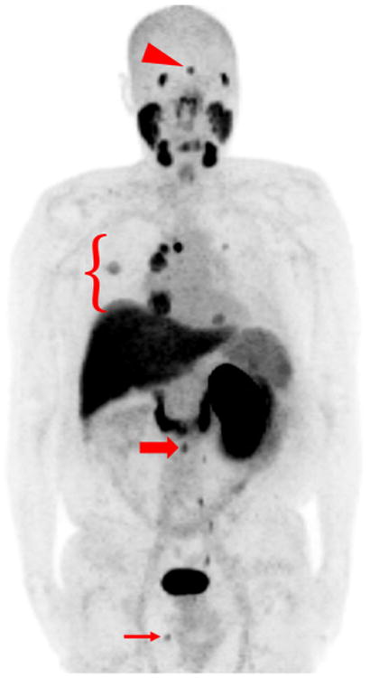
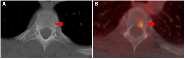
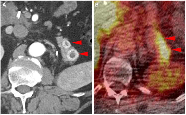
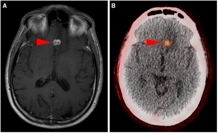
Results: In all five patients imaged, sites of putative metastatic disease were readily identifiable by abnormal ¹⁸F-DCFPyL uptake, with overall more lesions detected than on conventional imaging. These PET-detected sites included lymph nodes, pancreatic parenchymal lesions, lung parenchymal lesions, a brain parenchymal lesion, and other soft tissue sites. ¹⁸F-DCFPyL uptake ranged from subtle to intense with maximum standardized uptake values (SUVmax) for the identified lesions of 1.6-19.3. Based upon this small patient series, limited pathology and imaging follow-up of these patients suggests a higher sensitivity for ¹⁸F-DCFPyL compared to conventional imaging in the detection of metastatic RCC (94.7 versus 78.9%).
Conclusions: PSMA expression in the tumor neovasculature of RCC has been previously established and is believed to provide the basis for the imaging findings presented here. PSMA-based PET/CT with radiotracers such as ¹⁸F-DCFPyL may allow more accurate staging of patients with RCC and conceivably the ability to predict and follow therapy in patients treated with agents targeting the neovasculature.
Keywords: DCFPyL; Positron emission tomography (PET); Prostate-specific membrane antigen (PSMA); Renal cell carcinoma (RCC).
Conflict of interest statement
Figures


References
-
- Siegel RL, Miller KD, Jemal A. Cancer statistics, 2015. CA Cancer J Clin. 2015;65:5–29. - PubMed
-
- Brufau BP, Cerqueda CS, Villalba LB, Izquierdo RS, González BM, Molina CN. Metastatic renal cell carcinoma: radiologic findings and assessment of response to targeted therapy by using multidetector CT. Radiographics. 2013;33:1691–716. - PubMed
-
- Calderella C, Muoio B, Isgrò MA, Porfiri E, Treglia G, Gio-vanella L. The role of fluorine-18-fluorodeoxyglucose positron emission tomography in evaluating the response to tyrosine-ki-nase inhibitors in patients with metastatic primary renal cell carcinoma. Radiol Oncol. 2014;48:219–27. - PMC - PubMed
Publication types
MeSH terms
Substances
Grants and funding
LinkOut - more resources
Full Text Sources
Other Literature Sources
Medical
Research Materials
Miscellaneous
