. 2015 Aug 19:6:8088.
doi: 10.1038/ncomms9088.

Liquid demixing of intrinsically disordered proteins is seeded by poly(ADP-ribose)

Affiliations

Liquid demixing of intrinsically disordered proteins is seeded by poly(ADP-ribose)

Matthias Altmeyer et al. Nat Commun. .

Abstract

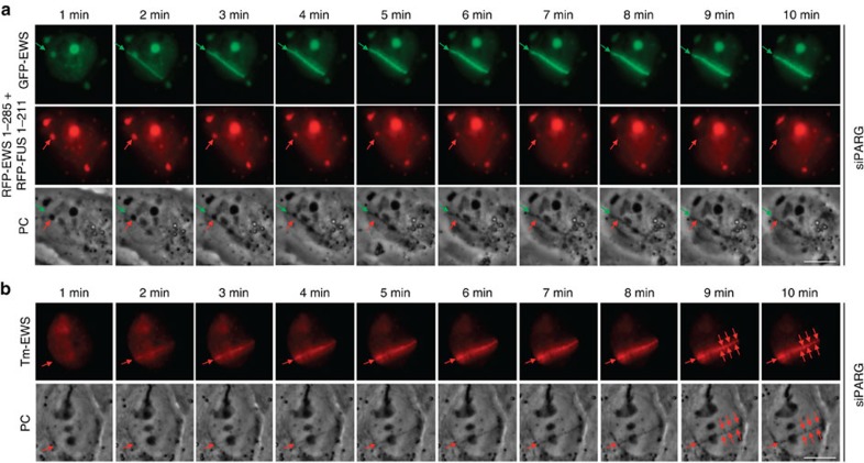
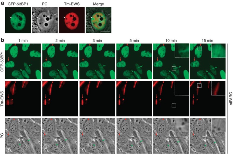
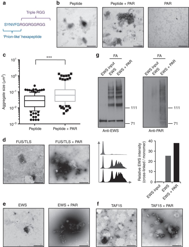
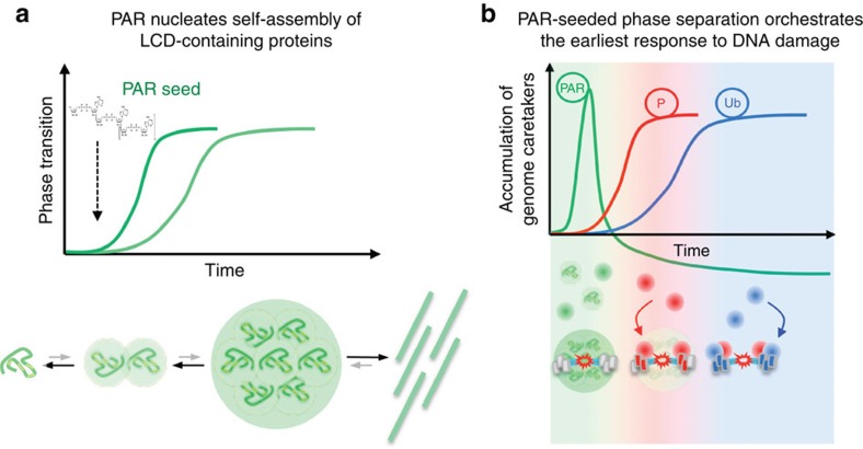
Intrinsically disordered proteins can phase separate from the soluble intracellular space, and tend to aggregate under pathological conditions. The physiological functions and molecular triggers of liquid demixing by phase separation are not well understood. Here we show in vitro and in vivo that the nucleic acid-mimicking biopolymer poly(ADP-ribose) (PAR) nucleates intracellular liquid demixing. PAR levels are markedly induced at sites of DNA damage, and we provide evidence that PAR-seeded liquid demixing results in rapid, yet transient and fully reversible assembly of various intrinsically disordered proteins at DNA break sites. Demixing, which relies on electrostatic interactions between positively charged RGG repeats and negatively charged PAR, is amplified by aggregation-prone prion-like domains, and orchestrates the earliest cellular responses to DNA breakage. We propose that PAR-seeded liquid demixing is a general mechanism to dynamically reorganize the soluble nuclear space with implications for pathological protein aggregation caused by derailed phase separation.


Figures

Figure 1
Figure 1. Intrinsically disordered proteins accumulate at sites of DNA damage in a PAR-dependent manner.
(a) Overlap of proteins associated with RNA granules (blue), b-isox precipitates and in vitro generated hydrogels (orange), and PAR (green). (b) Overlap of proteins associated with PAR (green) and a control group of 225 nuclear kinases (brown). (a,b) Asterisks indicate right-tailed P values (Fisher's exact test). (c) Protein domain organization of the LCD-containing FET proteins FUS, EWS and TAF15, each harbouring prion-like SYQG-rich amino termini and carboxyl termini rich in RGG repeats. Asterisks indicate regions of oncogenic translocations. (d) Recruitment kinetics of GFP–FUS to sites of laser microirradiation in the absence or presence of PARP inhibitor olaparib (10 μM). Time-lapse movie stills from the first 15 min after irradiation are shown. White arrows indicate the orientation of the laser line. See also Supplementary Movie 1. (e) Recruitment kinetics of GFP–EWS to sites of laser microirradiation. (f) Recruitment kinetics of GFP–TAF15 to sites of laser microirradiation. Scale bars, 10 μm.
Figure 2
Figure 2. The RGG modules in LCD-containing proteins function as sensors of PAR formation.
(a) Recruitment kinetics of GFP–TAF15 320–592 (20 RGGs) to sites of laser microirradiation in the absence or presence of PARP inhibitor olaparib (10 μM). Time-lapse movie stills from the first 15 min after irradiation are shown. White arrows indicate the orientation of the laser line. (b) Recruitment kinetics of GFP–EWS 445–656 (16 RGGs) to sites of laser microirradiation. (c) Recruitment kinetics of GFP–FUS 468–526 (8 RGGs) to sites of laser microirradiation. (d) GFP–FUS 468–526 in which RG/RGGs were altered to SG/SGG was expressed; cells were laser microirradiated and imaged as in ac. Scale bars, 10 μm.
Figure 3
Figure 3. Prion-like domains of LCD-containing proteins phase separate to form homotypic and heterotypic droplets by liquid demixing.
(a) GFP–FUS 1–211 was expressed for 24 h in U-2-OS cells and spontaneous intracellular droplet formation was detected by fluorescence and phase-contrast (PC) microscopy. (b) GFP–EWS 1–285 was expressed and analysed as in a. (c) GFP–TAF15 1–216 was expressed and analysed as in a. (d) Sub-nuclear formation of GFP–FUS 1–211 droplets, as scored and quantified by software-assisted image analysis using the Olympus ScanR system, is depicted as a function of GFP–FUS 1–211 expression to reveal a sharp transition towards droplet formation after reaching intra-nuclear concentrations sufficient to trigger spontaneous liquid demixing. (e) Sub-nuclear formation of GFP–EWS 1–285 was scored and analysed as in d. (f) Sub-nuclear formation of GFP–TAF15 1–216 was scored and analysed as in d. (g) GFP–FUS 1–211 was expressed as in a and monitored live by time-lapse imaging over a period of 1 h in 2-min intervals. Movie stills and magnifications of intracellular liquid droplets are provided. White asterisks mark a fusion event of two droplets, while red asterisks mark a fission event. See also Supplementary Movie 2. (h) GFP–FUS 1–211 and Tomato-EWS 1–285 were co-expressed for 24 h in U-2-OS cells and spontaneous formation of heterotypical droplets was detected by fluorescence and phase-contrast (PC) microscopy. (i) GFP–FUS 1–211 was expressed as in a, cells were fixed and stained for endogenous FUS using an antibody against the carboxyl terminus of the protein. (j) GFP–FUS 1–211 was expressed as in a, cells were fixed and stained for endogenous TAF15. (k) GFP–FUS 1–211 and Tomato-EWS 1–285 were co-expressed as in h, cells were fixed and stained for endogenous hnRNPUL1. (l) Tm-EWS 1–285 was expressed for 24 h, cells were laser microirradiated and imaged as in Fig. 2a–d. Time-lapse movie stills from the first 15 min after irradiation are shown. White arrows indicate the orientation of the laser line. (m) Tm-EWS 1–285 and GFP–EWS were co-expressed for 24 h, cells were laser microirradiated and imaged as in Fig. 2a–d. Time-lapse movie stills from the first 15 min after irradiation are shown. Scale bars, 10 μm.
Figure 4
Figure 4. PAR-dependent accumulation of LCD-containing proteins seeds liquid demixing at sites of DNA damage.
(a) Bright-field images depicting the transient generation of distinct light-diffracting stripes at sites of laser microirradiation under conditions of increased laser energy. White arrows indicate the orientation of the laser line, black arrows point to light-diffracting stripes. Asterisk and dashed lines point to light-diffracting nucleoli. See Supplementary Materials for details. (b) Bright-field images depicting the enhanced and prolonged generation of distinct light-diffracting stripes at sites of laser microirradiation in PARG-depleted cells. See also Supplementary Movie 3. (c) Bright-field images of laser microirradiated PARG-depleted cells in the presence of PARP inhibitor. (d) Bright-field images of laser microirradiated siPARG/FET cells. (e) Transient ectopic expression of GFP–EWS enhances the generation of light-diffracting stripes in otherwise naive U-2-OS cells. Following laser microirradiation, cells were fixed and stained for PAR. White arrows in be indicate the orientation of the laser line, black arrows point to light-diffracting stripes. Scale bars, 10 μm.
Figure 5
Figure 5. Liquid demixing of LCD-containing proteins is dynamic and phase separated compartments can exchange its constituents.
(a) PARG-depleted U-2-OS cells were co-transfected with full-length GFP–EWS and the prion-like domains of EWS and FUS fused to RFP. Cells were laser microirradiated and time-lapse movie stills from the first 10 min after irradiation shown. Green arrows (upper panels) point to the recruitment of full-length GFP–EWS to DNA damage sites. Red arrows (middle panels) point to the redistribution of a prion-like domain-containing liquid droplet in the vicinity of the laser track. Note that the appearance of the distinct light-diffracting stripe at the laser microirradiated region is concomitant with the dissolution of the light-diffracting liquid droplet formed by the prion-like domains (lower panels). Prion domain containing droplets in distal regions of the nucleus and in the cytoplasm appeared stable during the period of observation. (b) PARG-depleted U-2-OS cells were transfected with full-length Tm-EWS. Cells were laser microirradiated and time-lapse movie stills from the first 10 min after irradiation shown. Red arrows (upper panels) point to the recruitment of Tm-EWS to DNA damage sites. Note that the full-length protein Tm-EWS dissolves into microdroplets at later time points. Scale bars, 10 μm.
Figure 6
Figure 6. PAR-initiated liquid demixing can filter protein interactions at damaged chromatin.
(a) Phase-contrast and fluorescent images depicting that sub-nuclear accumulation of the genome caretaker GFP–53BP1 correlates with decreased light diffraction and reduced levels of Tm-EWS (white arrows), while accumulation of Tm-EWS correlates with increased light diffraction and reduced levels of GFP–53BP1 (black arrows). (b) Movie snapshots from laser microirradiation experiments of GFP-53BP1/Tm-EWS co-expressing cells depicting reduced GFP–53BP1 accumulation at sites of EWS accumulation upon prolonged liquid demixing in PARG-depleted cells. Red arrows point at Tm-EWS accumulation, green arrows point at GFP–53BP1 accumulation. Scale bars, 10 μm.
Figure 7
Figure 7. Isolated PAR chains accelerate LCD aggregation in a cell-free system.
(a) Model peptide sequence designed to analyse PAR-seeded aggregation in vitro. The model peptide comprises a prion-like hexapeptide sequence followed by the three consecutive RGG repeats. (b) The model peptide was incubated at 37 °C for 24 h with or without sub-stoichiometric amounts of isolated, polydispersed PAR chains and spontaneous aggregates were analysed by transmission electron microscopy (TEM). (c) As in b, the model peptide was incubated with or without PAR, and aggregate sizes were determined from TEM images (n=137 for the peptide sample; n=116 for the peptide+PAR sample). ***P<0.0001 (Mann–Whitney test). (d) Full-length recombinant FUS was incubated at 37 °C for 24 h with or without sub-stoichiometric amounts of purified PAR and protein aggregates were analysed by TEM. (e) Full-length recombinant EWS was incubated at 37 °C for 24 h with or without sub-stoichiometric amounts of purified PAR and protein aggregates were analysed by TEM. (f) Full-length recombinant TAF15 was incubated at 37 °C for 24 h with or without sub-stoichiometric amounts of purified PAR and protein aggregates were analysed by TEM. All TEM experiments were repeated at least three times, and representative images are shown. Additional images are provided as Supplementary Fig. 8. Scale bars, 500 nm. (g) Full-length recombinant EWS was incubated with or without purified PAR, cross-linked in 0.4% formaldehyde (FA) for 15 min, and analysed by SDS–polyacrylamide gel electrophoresis (3–8% Tris–acetate). After detection of EWS complexes (left panel), the membrane was stripped and reprobed with an antibody against PAR (right panel). Signals from the anti-EWS western blot were quantified by ImageJ.
Figure 8
Figure 8. Model for PAR-nucleated liquid demixing of LCD-containing proteins.
(a) Intrinsically disordered LCD-containing proteins undergo spontaneous self-assembly to generate higher-order structures. The initial kinetics during the nucleation phase of this process is relatively slow due to fast reverse reaction rates. However, molecular seeds can significantly accelerate the nucleation process, thereby help to overcome the kinetic barrier and drive the formation of higher-order structures; in case of excessive or lasting stimuli pathological fibrils or irreversible protein aggregates may form. We propose that the low complexity anionic biopolymer poly(ADP-ribose) (PAR) constitutes a molecular seed for the self-assembly of LCD-containing proteins. By virtue of its non-rigid structure and polydispersed nature, PAR can trap intrinsically disordered LCD-containing proteins and facilitate their dynamic assembly into higher-order structures. Under physiological conditions, the PAR-seeded assembly of LCD-containing proteins thus represents a liquid–liquid phase separation, with the potential to dynamically compartmentalize the subcellular space in a context-dependent manner. Under pathological conditions, derailed phase transitions may lead to the formation of less dynamic protein aggregates. (b) In the physiological context of the cellular response to DNA damage, PAR levels spike locally due to hyperactivation of PARP enzymes directly at DNA break sites, resulting in the rapid accumulation of various LCD-containing proteins. Accordingly, the greatly increased local concentration of LCD-containing proteins results in rapid phase separation and liquid demixing, providing cells with an opportunity to filter molecular interactions occurring on damaged chromatin. Dissolution of PAR-seeded liquid compartments paves the way for dedicated high-affinity key–lock interactions to unfold on the lesion-flanking chromatin allowing for the accumulation of genome caretakers such as 53BP1.

Comment in

Similar articles

Cited by

References

    1. Malinovska L., Kroschwald S. & Alberti S. Protein disorder, prion propensities, and self-organizing macromolecular collectives. Biochim. Biophys. Acta. 1834, 918–931 (2013). - PubMed
    1. Li Y. R., King O. D., Shorter J. & Gitler A. D. Stress granules as crucibles of ALS pathogenesis. J. Cell Biol. 201, 361–372 (2013). - PMC - PubMed
    1. Jucker M. & Walker L. C. Self-propagation of pathogenic protein aggregates in neurodegenerative diseases. Nature 501, 45–51 (2013). - PMC - PubMed
    1. Han T. W. et al.. Cell-free formation of RNA granules: bound RNAs identify features and components of cellular assemblies. Cell 149, 768–779 (2012). - PubMed
    1. Kato M. et al.. Cell-free formation of RNA granules: low complexity sequence domains form dynamic fibers within hydrogels. Cell 149, 753–767 (2012). - PMC - PubMed

Publication types