Age differences in hippocampal subfield volumes from childhood to late adulthood
- PMID: 26286891
- PMCID: PMC4718822
- DOI: 10.1002/hipo.22517
Age differences in hippocampal subfield volumes from childhood to late adulthood
Abstract
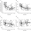
The hippocampus is composed of distinct subfields: the four cornu ammonis areas (CA1-CA4), dentate gyrus (DG), and subiculum. The few in vivo studies of human hippocampal subfields suggest that the extent of age differences in volume varies across subfields during healthy childhood development and aging. However, the associations between age and subfield volumes across the entire lifespan are unknown. Here, we used a high-resolution imaging technique and manually measured hippocampal subfield and entorhinal cortex volumes in a healthy lifespan sample (N = 202), ages 8-82 yrs. The magnitude of age differences in volume varied among the regions. Combined CA1-2 volume evidenced a negative linear association with age. In contrast, the associations between age and volumes of CA3-DG and the entorhinal cortex were negative in mid-childhood and attenuated in later adulthood. Volume of the subiculum was unrelated to age. The different magnitudes and patterns of age differences in subfield volumes may reflect dynamic microstructural factors and have implications for cognitive functions across the lifespan. © 2015 Wiley Periodicals, Inc.
Keywords: CA1; aging; dentate gyrus; development; entorhinal cortex.
© 2015 Wiley Periodicals, Inc.
Figures


References
-
- Amaral D, Lavenex P. Hippocampal neuroanatomy. In: Andersen P, Morris R, Amaral D, Bliss T, O'Keefe J, editors. The Hippocampus Book Oxford Neuroscience Series. New York: Oxford University Press; 2006. pp. 37–107.
-
- Bender AR, Daugherty AM, Raz N. Vascular risk moderates associations between hippocampal subfield volumes and memory. J Cog Neuro. 2013;25(1):1851–1862. - PubMed
-
- Bobinski M, de Leon MJ, Wegiel J, Desanti S, Convit A, Saint Louis LA, Rusinek H, Wisniewski HM. The histological validation of post mortem magnetic resonance imaging-determined hippocampal volume in Alzheimer's disease. Neurosci. 2000;95(3):721–725. - PubMed
-
- Breunig JJ, Arellano JI, Macklis JD, Rakic P. Everything that glitters isn’t gold: A critical review of postnatal neural precursor analyses. Cell Stem Cell. 2007;1:612–627. - PubMed
Publication types
MeSH terms
Grants and funding
LinkOut - more resources
Full Text Sources
Other Literature Sources
Medical
Miscellaneous

