

Porous Silicon Structures as Optical Gas Sensors
- PMID: 26287199
- PMCID: PMC4570405
- DOI: 10.3390/s150819968
Porous Silicon Structures as Optical Gas Sensors
Abstract
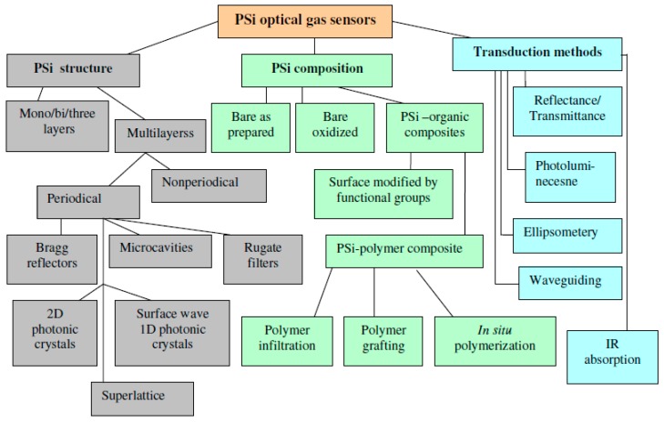
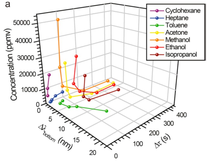
We present a short review of recent progress in the field of optical gas sensors based on porous silicon (PSi) and PSi composites, which are separate from PSi optochemical and biological sensors for a liquid medium. Different periodical and nonperiodical PSi photonic structures (bares, modified by functional groups or infiltrated with sensory polymers) are described for gas sensing with an emphasis on the device specificity, sensitivity and stability to the environment. Special attention is paid to multiparametric sensing and sensor array platforms as effective trends for the improvement of analyte classification and quantification. Mechanisms of gas physical and chemical sorption inside PSi mesopores and pores of PSi functional composites are discussed.
Keywords: gas sensors; multiparametric; optical; porous silicon; sensor arrays.
Figures
Similar articles
-
Optical microsensors for pesticides identification based on porous silicon technology.Biosens Bioelectron. 2005 Apr 15;20(10):2136-9. doi: 10.1016/j.bios.2004.09.020. Biosens Bioelectron. 2005. PMID: 15741087
-
Periodically porous top electrodes on vertical nanowire arrays for highly sensitive gas detection.Nanotechnology. 2011 Sep 2;22(35):355501. doi: 10.1088/0957-4484/22/35/355501. Epub 2011 Aug 5. Nanotechnology. 2011. PMID: 21817785
-
Silicon Photonic Biosensors Using Label-Free Detection.Sensors (Basel). 2018 Oct 18;18(10):3519. doi: 10.3390/s18103519. Sensors (Basel). 2018. PMID: 30340405 Free PMC article. Review.
-
Two-dimensional wavelet transform feature extraction for porous silicon chemical sensors.Anal Chim Acta. 2013 Jun 27;785:1-15. doi: 10.1016/j.aca.2013.04.024. Epub 2013 Apr 18. Anal Chim Acta. 2013. PMID: 23764437
-
Porous Silicon Optical Biosensors: Still a Promise or a Failure?Sensors (Basel). 2019 Nov 3;19(21):4776. doi: 10.3390/s19214776. Sensors (Basel). 2019. PMID: 31684128 Free PMC article. Review.
Cited by
-
Nanoscale morphology, optical dynamics and gas sensor of porous silicon.Sci Rep. 2024 Feb 14;14(1):3677. doi: 10.1038/s41598-024-54336-x. Sci Rep. 2024. PMID: 38355956 Free PMC article.
-
Ultra-high sensitive 1D porous silicon photonic crystal sensor based on the coupling of Tamm/Fano resonances in the mid-infrared region.Sci Rep. 2019 May 6;9(1):6973. doi: 10.1038/s41598-019-43440-y. Sci Rep. 2019. PMID: 31061422 Free PMC article.
-
A Pyrazine-Based Chromophore Photophysical Properties and its Detection for Hydrazine and Acid Vapors.J Fluoresc. 2025 Jun;35(6):4039-4051. doi: 10.1007/s10895-024-03825-3. Epub 2024 Jul 1. J Fluoresc. 2025. PMID: 38951307
-
A Robust Distributed Multipoint Fiber Optic Gas Sensor System Based on AGC Amplifier Structure.Sensors (Basel). 2016 Jul 28;16(8):1187. doi: 10.3390/s16081187. Sensors (Basel). 2016. PMID: 27483267 Free PMC article.
-
Quasi periodic photonic crystal as gamma detector using Poly nanocomposite and porous silicon.Sci Rep. 2025 May 27;15(1):18451. doi: 10.1038/s41598-025-02910-2. Sci Rep. 2025. PMID: 40419577 Free PMC article.
References
-
- Sailor M.J. Porous Silicon in Practice. Wiley-VCH Verlag; Weinheim, Germany: 2012.
-
- Lauerhaas J.M., Gredo G.M., Heinrich J.L., Sailor M.J. Reversible luminescence quenching of porous Si by solvents. J. Am. Chem. Soc. 1992;114:1911–1912. doi: 10.1021/ja00031a072. - DOI
-
- Harraz F.A. Porous silicon chemical sensors and biosensors: A review. Sens. Actuators B Chem. 2014;202:897–912. doi: 10.1016/j.snb.2014.06.048. - DOI
Publication types
MeSH terms
Substances
LinkOut - more resources
Full Text Sources
Other Literature Sources
