. 2015 Sep 17;525(7569):389-393.
doi: 10.1038/nature15252. Epub 2015 Aug 19.

Tet2 is required to resolve inflammation by recruiting Hdac2 to specifically repress IL-6

Affiliations

Tet2 is required to resolve inflammation by recruiting Hdac2 to specifically repress IL-6

Qian Zhang et al. Nature. .

Abstract

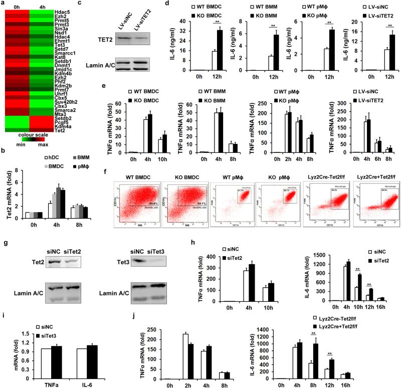
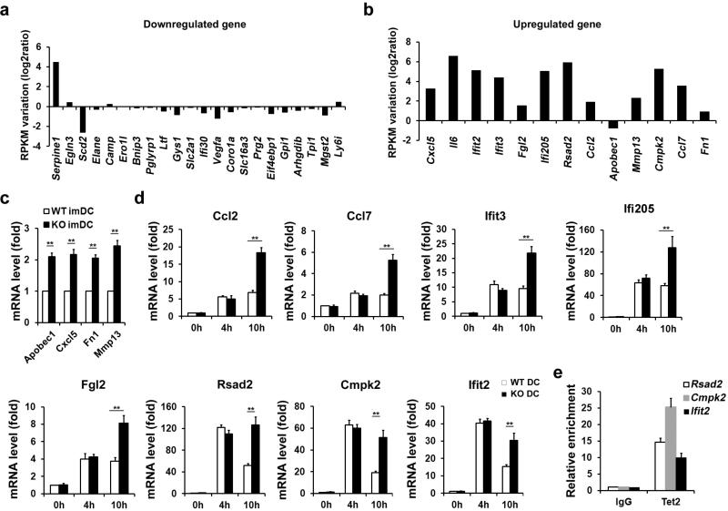
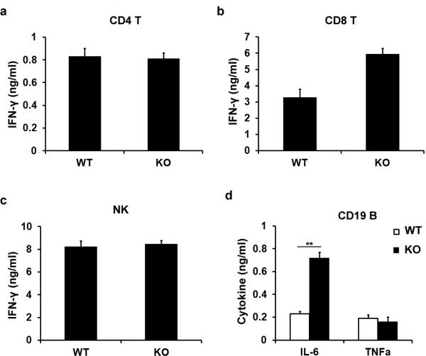
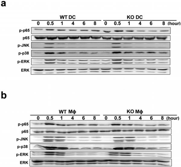
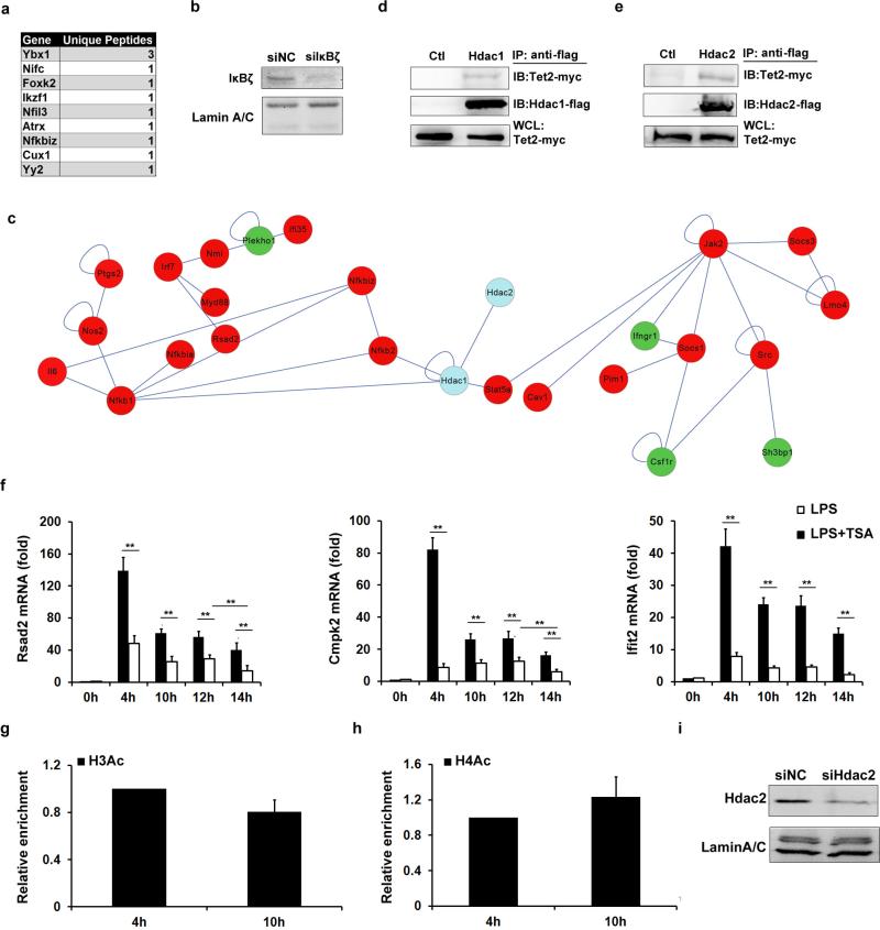
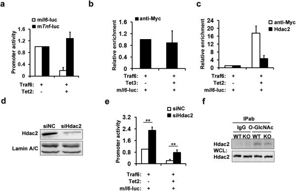
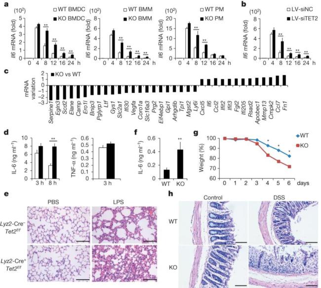
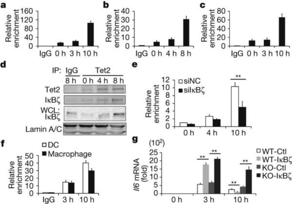
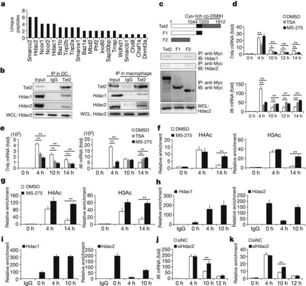
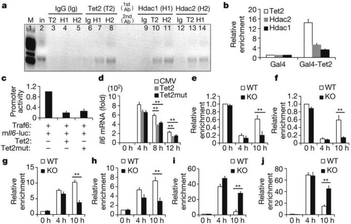
Epigenetic modifiers have fundamental roles in defining unique cellular identity through the establishment and maintenance of lineage-specific chromatin and methylation status. Several DNA modifications such as 5-hydroxymethylcytosine (5hmC) are catalysed by the ten eleven translocation (Tet) methylcytosine dioxygenase family members, and the roles of Tet proteins in regulating chromatin architecture and gene transcription independently of DNA methylation have been gradually uncovered. However, the regulation of immunity and inflammation by Tet proteins independent of their role in modulating DNA methylation remains largely unknown. Here we show that Tet2 selectively mediates active repression of interleukin-6 (IL-6) transcription during inflammation resolution in innate myeloid cells, including dendritic cells and macrophages. Loss of Tet2 resulted in the upregulation of several inflammatory mediators, including IL-6, at late phase during the response to lipopolysaccharide challenge. Tet2-deficient mice were more susceptible to endotoxin shock and dextran-sulfate-sodium-induced colitis, displaying a more severe inflammatory phenotype and increased IL-6 production compared to wild-type mice. IκBζ, an IL-6-specific transcription factor, mediated specific targeting of Tet2 to the Il6 promoter, further indicating opposite regulatory roles of IκBζ at initial and resolution phases of inflammation. For the repression mechanism, independent of DNA methylation and hydroxymethylation, Tet2 recruited Hdac2 and repressed transcription of Il6 via histone deacetylation. We provide mechanistic evidence for the gene-specific transcription repression activity of Tet2 via histone deacetylation and for the prevention of constant transcription activation at the chromatin level for resolving inflammation.


Figures

Extended Data Figure 1
Extended Data Figure 1. Loss of Tet2 specifically enhances expression of IL-6 but not TNF-α
a, Hierarchical cluster of transcription level of chromatin modifiers with significant variation in LPS-stimulated murine BMDC, RPKMs were centred by the mean. b,Tet2 mRNA in human dendritic cells (hDC), LPS-stimulated murine BMDC, BMM and primary peritoneal macrophages (pMϕ). cf, j, Human imDCs in which TET2 were lentivirally silenced (LV-siTET2), BMDC, BMM and peritoneal macrophages from Tet2-deficient mice (cf), peritoneal macrophages from conditional Tet2-deficient mice (f, j) and their respective controls were stimulated by LPS for 8 h (c) or the indicated time (d, e, j). Protein levels of TET2 were detected by immunoblot, with lamin A/C as the loading control (c). IL-6 protein levels were analysed by ELISA (d), and Tnfa and Il6 mRNA levels were analysed by qPCR (e, j). Percentage of Iab+CD11c+BMDC or F4/80+ CD11b+peritoneal macrophages were analysed (f). gi, Tet2 (g, h), Tet3 (g, i) were silenced in peritoneal macrophages for 48 h and then peritoneal macrophages were stimulated with LPS for 8 h (g, i) or the indicated time (h), and the protein levels of Tet2 or Tet3 were detected by immunoblot, with lamin A/C as the loading control (g). Tnfa and Il6 mRNA were analysed by qPCR (h, i). Full scans of blots are shown in Supplementary Fig. 1. Error bars represent s.e.m. of triplicate biological replicates and are representative of three independent experiments. **P < 0.01.
Extended Data Figure 2
Extended Data Figure 2. Loss of Tet2 barely affects development of immune cells
Percentages of the indicated immune cells were analysed by their specific lineage markers from control and Tet2-deficient mice. Data were from one representative of three independent experiments.
Extended Data Figure 3
Extended Data Figure 3. Gene expression variation in Tet2-deficient dendritic cells
a, b, mRNA variations of indicated genes in RNA-seq analysis of BMDC stimulated with LPS. RPKM of each of the genes in 4 h group were compared with 0 h group, and calculated to log2 ratio. c, d, qPCR analysis of mRNA levels of indicated genes in wild type and Tet2-deficient naive BMDC (c) or LPS-stimulated BMDC (d). e, BMDC were stimulated with LPS for 8 h, and then endogenous Tet2 enrichments at proximal promoter of indicated genes were analysed by ChIP-qPCR. Error bars represent s.d. of triplicate technical (e) or s.e.m. of triplicate biological (c, d) replicates and are representative of three independent experiments. **P < 0.01.
Extended Data Figure 4
Extended Data Figure 4. Potential targets of Tet2 during immune response
ad, ELISA of cytokines in supernatants of splenic activated CD4+ T cells (a), CD8+ T cells (b) and NK cells (c), and conventional splenic CD19+ B cells (d) from Tet2-deficient (KO) and control mice (WT). T cells were stimulated with 10 μg ml−1 anti-CD3 and 1 μg ml−1 anti-CD28 monoclonal antibodies for 24 h. NK cells were stimulated with a combination of 10 ng ml−1 IL-12 plus 5 ng ml−1 IL-18 for 12 h. B cells were stimulated with 5 μg ml−1 LPS for 24 h, Error bars represent s.d. of triplicate technical replicates and are representative of three independent experiments. **P < 0.01.
Extended Data Figure 5
Extended Data Figure 5. Loss of Tet2 barely affects TLR4 signalling pathways
a, b, Immunoblot assays of the phosphorylated (p-) or total proteins in lysates of wild-type and Tet2-deficent BMDC (a) and peritoneal macrophages (b) stimulated with LPS for indicated time. Full scans of blots are shown in Supplementary Fig. 1. Data were from one representative of three independent experiments.
Extended Data Figure 6
Extended Data Figure 6. Tet2 binding partners for cytokine regulation
a, Co-immunoprecipitation assay using Tet2 antibody of nuclear fraction of murine BMDC stimulated with LPS for 8 h. Gene symbols of transcription factor deposited in KEGG database and their unique peptides identified by mass spectrometry analysis were tabled. b, IκBζ was silenced in BMDC using specific siRNA for 36 h and the BMDC were stimulated with LPS for 8 h. The protein levels of IκBζ were detected by immunoblot, with lamin A/C as the loading control. c, Hdac1/2 (blue circles) were subjected to interaction analysis with genes which had significant expression variations (red circles for upregulated, and green circles for downregulated) in BMDC 4 h after LPS stimulation. The linkers indicate interaction or regulation relationship between the two genes. d, e, Flag-tagged Hdac1 (d) or Hdac2 (e) were overexpressed in HEK293T cells together with Myc-tagged Tet2. Cell lysates were examined by IP and immunoblot with indicated antibodies. The whole-cell lysates (WCL) were used to examine the input of overexpressed proteins. f, BMDC were pretreated with 100 nM TSA for 1 h, and then stimulated by LPS for indicated time. mRNA levels of indicated genes were analysed by qPCR. g, h, BMDC (g) and peritoneal macrophages (h) were stimulated with LPS for the indicated time. Enrichments of H3Ac (g), H4Ac (h) at Tnf promoters were analysed by ChIP-qPCR. i, Hdac2 was silenced in BMDC for 48 h and then the BMDC were stimulated with LPS for 8 h. The protein levels of Hdac2 were detected by immunoblot, with lamin A/C as the loading control. Full scans of blots are shown in Supplementary Fig. 1. Error bars represent s.d. of triplicate technical (g, h) or s.e.m. of triplicate biological (f) replicates and are representative of three independent experiments. **P < 0.01.
Extended Data Figure 7
Extended Data Figure 7. Tet2 represses Il6 promoter activity through recruiting Hdac2
a, Luciferase activities in lysates of HEK293T cells transfected with indicated plasmids and luciferase reporter for murine Il6 promoter (TSS to upstream 3K). b, ChIP-qPCR analysis of Myc-tagged Tet3 in murine Il6 promoter in HEK293T cells which were transfected with indicated plasmids and luciferase reporter. c, ChIP-qPCR of Myc-tagged Tet2 or endogenous Hdac2 in murine Il6 promoter in HEK293T cells transfected with indicated plasmids and luciferase reporter. d, Hdac2 was silenced for 36 h in HEK293T cells. Protein levels of Hdac2 were detected by immunoblot, with lamin A/C as the loading control. e, Luciferase activities in lysates of Hdac2-silenced HEK293T cells transfected with indicated plasmids and luciferase reporter containing murine Il6 promoter. f, Wild-type (WT) and Tet2-deficient (KO) peritoneal macrophages were stimulated with LPS for 8 h. IP using O-GlcNAcylation antibody was performed. The whole-cell lysates (WCL) were used as the input. Full scans of blots are shown in Supplementary Fig. 1. Error bars represent s.d. of triplicate technical replicates and are representative of three independent experiments. **P < 0.01.
Figure 1
Figure 1. Loss of Tet2 maintains higher expression of IL-6
a, b, Il6 mRNA in BMDC, bone-marrow-derived macrophages (BMM) and peritoneal macrophages (PM) (a) from wild-type (WT) and Tet2-knockout (KO) mice, and TET2-silenced human dendritic cells (b) during LPS response. c, log2 ratio of mRNA variations in Tet2-deficient BMDC 8 h after LPS stimulation. d, e, ELISA of sera cytokines (d) and histopathology of lungs (e) from conditional Tet2-deficient and control mice (n = 5) after intra-peritoneal injection of LPS (10 mg per kg body weight). Scale bars, 50 μm. f, g, h, ELISA of sera IL-6 (f), changes of body weights (g) and histopathology of colonic sections (h) of Tet2-deficient and control mice (n = 5) on day 6 after treatment with 3% DSS. Scale bars, 100 μm. Error bars represent s.d. of triplicate technical replicates (b, d, f) or s.e.m. of triplicate biological replicates (a). Data are representative of 3 independent experiments. Unpaired Student's t-test, *P < 0.05, **P < 0.01.
Figure 2
Figure 2. IκBζ mediates selective targeting of Tet2 to Il6 promoter
ac, e, f, ChIP followed by quantitative PCR (ChIP-qPCR) of Tet2 (ac, e) and IκBζ (f) at the Il6 promoter in BMDC (a, f), peritoneal macrophages (b, f) and human dendritic cells (c) and IκBζ-silenced BMDC (e). IP, immunoprecipitation. d, Tet2 interacted with IκBζ in LPS-stimulated BMDC. g, Il6 mRNA in IκBζ-overexpressed wild-type and Tet2-deficient peritoneal macrophages. Cells were stimulated by LPS. Full scans of blots are shown in Supplementary Fig. 1. Error bars represent s.d. of triplicate technical replicates. Data are representative of 3 independent experiments. Unpaired Student's t-test, **P < 0.01.
Figure 3
Figure 3. Hdac2 associates with Tet2 and specifically represses IL-6 through histone deacetylation
a, MS-identified Tet2-binding chromatin regulators in LPS-stimulated BMDC for 8 h. b,Tet2 interacts with HDAC1/2 in cells 8 h after LPS stimulation. c, Deletion mutants of Tet2 were overexpressed to detect endogenous HDAC1/2 association. IB, immunoblot. di, BMDC (d, f, h) and peritoneal macrophages (e, g, i) were pre-treated with 100 nM TSA or 4 μM MS-275 (dg), or left naive (h, i), then stimulated with LPS. Cytokine mRNA (d, e) and histone acetylation and Hdac1/2 at Il6 promoter (fi) were analysed. j, k, Il6 mRNA and H3Ac enrichment at Il6 promoter in Hdac2-silenced (siHdac2) BMDC stimulated with LPS. Full scans of blots are shown in Supplementary Fig. 1. Error bars represent s.d. of triplicate technical (fi, k) or s.e.m. of triplicate biological (d, e, j) replicates. Data are representative of 3 independent experiments. Unpaired Student's t-test, *P < 0.05, **P < 0.01.
Figure 4
Figure 4. Tet2 recruits Hdac2 to repress expression of IL-6
a, Re-ChIP-PCR assay of Tet2–Hdac1/2 interaction at Il6 promoter in BMDC 8 h after LPS stimulation. First round ChIP (1st antibody, Ab) against Tet2 (T2), Hdac1 (H1), Hdac2 (H2) or IgG (Ig). Eluted samples were re-ChIPed (2nd antibody). Lane 2 was input (in). b, ChIP-qPCR assay of Tet2 and Hdac1/2 at Gal4-binding sites in Gal4-Tet2-overexpressing peritoneal macrophages. c, Luciferase activities in lysates of HEK293T cells transfected with indicated plasmids and murine Il6 promoter reporter (mIl6-luc). d, Il6 mRNA in wild-type Tet2- or catalytic mutant Tet2-overexpressed Tet2-deficient peritoneal macrophages stimulated with LPS. ej, Tet2-deficient BMDC (e, g, i) and peritoneal macrophages (f, h, j) were stimulated with LPS. Hdac2 (e, f), Hdac1(g, h), H3Ac (i), H4Ac (j) enrichments at Il6 promoter were analysed by ChIP-qPCR. Full scan of gel is shown in Supplementary Fig. 1. Error bars represent s.d. of triplicate technical replicates (b, dj) or s.e.m. of triplicate biological replicates (c). Data are representative of 3 independent experiments. Unpaired Student's t-test, **P < 0.01.

Comment in

  • Inflammation: TET2: the terminator.
    Bird L. Bird L. Nat Rev Immunol. 2015 Oct;15(10):598. doi: 10.1038/nri3912. Epub 2015 Sep 11. Nat Rev Immunol. 2015. PMID: 26358395 No abstract available.

References

    1. Chen T, Dent SY. Chromatin modifiers and remodellers: regulators of cellular differentiation. Nature Rev. Genet. 2014;15:93–106. - PMC - PubMed
    1. Kohli RM, Zhang Y. TET enzymes, TDG and the dynamics of DNA demethylation. Nature. 2013;502:472–479. - PMC - PubMed
    1. Pastor WA, Aravind L, Rao A. TETonic shift: biological roles of TET proteins in DNA demethylation and transcription. Nature Rev. Mol. Cell Biol. 2013;14:341–356. - PMC - PubMed
    1. Tahiliani M, et al. Conversion of 5-methylcytosine to 5-hydroxymethylcytosine in mammalian DNA by MLL partner TET1. Science. 2009;324:930–935. - PMC - PubMed
    1. Ko M, et al. Impaired hydroxylation of 5-methylcytosine in myeloid cancers with mutant TET2. Nature. 2010;468:839–843. - PMC - PubMed

Publication types

MeSH terms

Associated data