. 2015 Sep 15;6(27):23890-904.
doi: 10.18632/oncotarget.4341.

NCOA1 promotes angiogenesis in breast tumors by simultaneously enhancing both HIF1α- and AP-1-mediated VEGFa transcription

Affiliations

NCOA1 promotes angiogenesis in breast tumors by simultaneously enhancing both HIF1α- and AP-1-mediated VEGFa transcription

Li Qin et al. Oncotarget. .

Abstract

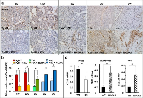
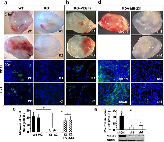
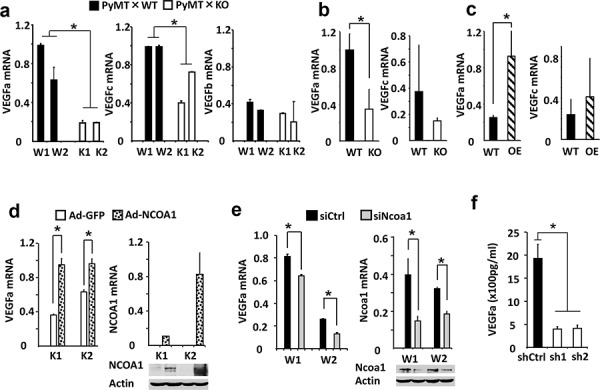
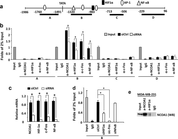
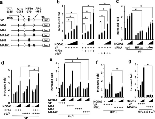
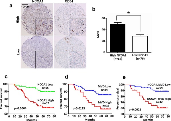
Nuclear receptor coactivator 1 (NCOA1) is overexpressed in a subset of breast cancer and its increased expression positively correlates with disease recurrence and metastasis. Although NCOA1 is known to promote breast cancer metastasis through working with multiple transcription factors to upregulate the expression of Twist1, ITGA5, CSF-1, SDF1 and CXCR4, the role of NCOA1 in breast tumor angiogenesis has not been investigated. In this study, we found that the microvascular density (MVD) was significantly decreased and increased in Ncoa1-knockout and NCOA1-overexpressing mammary tumors, respectively, in several breast cancer mouse models. Knockout or knockdown of NCOA1 in breast cancer cell lines also markedly compromised their capability to induce angiogenesis in Matrigel plugs embedded subcutaneously in mice, while this compromised capability could be rescued by VEGFa treatment. At the molecular level, NCOA1 upregulates VEGFa expression in both mouse mammary tumors and cultured breast cancer cells, and it does so by associating with both c-Fos, which is recruited to the AP-1 site at bp -938 of the VEGFa promoter, and HIF1α, which is recruited to the HIF1α-binding element at bp -979 of the VEGFa promoter, to enhance VEGFa transcription. In 140 human breast tumors, high NCOA1 protein correlates with high MVD and patients with both high NCOA1 and high MVD showed significantly shorter survival time. In summary, this study revealed a novel mechanism that NCOA1 potentiates breast cancer angiogenesis through upregulating HIF1α and AP-1-mediated VEGFa expression, which reinforces the rational of targeting NCOA1 in controlling breast cancer progression and metastasis.

Keywords: NCOA1; VEGFa; breast cancer; transcriptional regulation.


Conflict of interest statement

CONFLICTS OF INTEREST

The authors have no conflicting financial interests.

Figures

Figure 1
Figure 1. Microvascular density (MVD) in mouse mammary tumors with Ncoa1 knockout or overexpression
a. Detection of CD31-positive endothelial cells by immunohistochemistry in mouse mammary tumor tissue sections prepared from Tg(MMTV-PyMT), Tg(MMTV-PyMT) × Ncoa1−/−, Tg(MMTV-TVA/RCAS-PyMT), Tg(MMTV-TVA/RCAS-PyMT) × Tg(MMTV-NCOA1), Tg(MMTV-Neu) and Tg(MMTV-Neu) × Tg(MMTV-NCOA1) mice as indicated. Tumors were isolated from mice after palpable tumors were detected for the time in weeks indicated. Scale bar: 50 μm. b. Semi-quantitative analysis of MVD. The total number of microvessels in 5 different viewing fields of 200× magnification under a microscope was counted for each tumor section. Sections from at least 10 tumors in each group were examined. Data are presented as Mean ± SD. *p < 0.05 by Student's t test. c. QPCR analysis of CD31 mRNA in the mouse mammary tumors (n = 5) isolated from mice with the indicated genotypes. PyMT, Tg(MMTV-PyMT); TVA/PyMT, Tg(MMTV-TVA/RCAS-PyMT); Neu, Tg(MMTV-Neu); WT, wild type; NCOA1, Tg(MMTV-NCOA1).
Figure 2
Figure 2. In vivo Matrigel angiogenesis induced by mouse and human breast tumor cells with Ncoa1 knockout and NCOA1 knockdown, respectively
a. Representative images of Matrigel plugs with angiogenesis induced by W1, W2, K1 and K2 cells in SCID/beige mice (upper four panels) and representative images of CD31 and Flk1 immunofluorescent staining (green color) and DAPI staining (blue color) of Matrigel plug sections with angiogenesis induced by W1 and K1 cells (lower four panels). b. Representative images of Matrigel plugs with angiogenesis induced by K1 and K2 cells as well as 10 nM of recombinant VEGFa (rVEGFa) protein in Matrigel (upper two panels) and representative images of CD31 and Flk1 immunofluorescent staining and DAPI staining of Matrigel plug sections with angiogenesis induced by K1 cells plus rVEGFa (lower two panels). c. Semi-quantitative analysis of angiogenesis induced by W1, W2, K1 and K2 cells as well as K1 and K2 cells plus rVEGFa in Matrigel plugs in mice. A total of 12 Matrigel plugs with angiogenesis induced by the indicated cells with or without rVEGFa were analyzed. The number of microvessels per 200× viewing field was counted and 5 viewing fields were examined for each Matrigel plug. The data are presented as Mean ± SD. *p < 0.05 by Student's t test. d. Representative images of Matrigel plugs with angiogenesis induced by MDA-MB-231 cells expressing non-targeting shRNA (shCtrl) or NCOA1 mRNA-targeting shRNAs (sh1 and sh2) and representative images of CD31 and Flk1 immunofluorescent staining and DAPI staining of Matrigel plug sections with angiogenesis induced by MDA-MB-231 cells expressing shCtrl or sh1. e. Semi-quantitative analysis of angiogenesis induced by MDA-MB-231 cells expressing shCtrl, sh1 or sh2 in Matrigel plugs. Microvessels in 6 Matrigel plugs for each group were examined and counted as described above. Data are presented as Mean ± SD. *p < 0.05 by Student's t test. The knockdown efficiency of NCOA1 was analyzed by Western blot.
Figure 3
Figure 3. NCOA1 regulates VEGFa expression in breast tumor cells
a. Relative expression levels of VEGFa, VEGFc and VEGFb mRNAs in W1, W2, K1 and K2 cells measured by QPCR. b. Relative expression levels of VEGFa and VEGFc mRNAs in Tg(MMTV-PyMT) (WT) and Tg(MMTV-PyMT) × Ncoa1−/− (KO) mouse mammary tumors (n = 5) measured by QPCR. c. Relative expression levels of VEGFa and VEGFc mRNA levels in Tg(MMTV-TVA/RCAS-PyMT) (WT) and Tg(MMTV-TVA/RCAS-PyMT) × Tg(MMTV-NCOA1) (OE) mouse mammary tumors (n = 10) measured by QPCR. d. Relative expression levels of VEGFa mRNA in K1 and K2 cells with adenovirus-mediated GFP or NCOA1 expression (left panel). The expression levels of NCOA1 were analyzed by both QPCR and Western blotting (right panel). As expected, human NCOA1 mRNA was not expressed in the mouse K1 and K2 cells. e. Relative expression levels of VEGFa and Ncoa1 mRNAs in W1 and W2 cells transfected with siCtrl or Ncoa1 siRNAs as indicated. The measurement was carried out by QPCR. Ncoa1 knockdown efficiency was also analyzed by Western blotting. f. Secreted VEGFa concentrations in the conditioned media of MDA-MB-231 cells expressing non-targeting control shRNA (shCtrl) or NCOA1 mRNA-targeting shRNAs (sh1 and sh2). NCOA1 knockdown efficiency in these cells was shown in Figure 2e. The * in all panels indicates p < 0.05 by Student's t test.
Figure 4
Figure 4. NCOA1 is recruited to the VEGFa promoter by HIF1α and c-Fos
a. The VEGFa promoter region contains a TATA box and one HIF1a, three AP-1 and one NF-κB binding sites that are known to regulate VEGFa expression. Regions A–D were used for PCR amplification in ChIP assays. b. ChIP assays performed in MDA-MB-231 cells transfected with the non-targeting siRNA Smart Pool (siCtrl) or siRNA Smart Pools targeting NCOA1, HIF1α, c-Fos or NF-κB mRNAs as indicated. NCOA1, HIF1α, c-Fos and NF-κB antibodies were used for ChIP and normal IgG was used as negative control for ChIP. DNA obtained from ChIP was used as template for QPCR to measure the relative DNA amounts of regions A–D of the VEGFa promoter. The QPCR results for these regions were normalized to the QPCR results of 2% input DNA. c. The relative levels of HIF1α, c-Fos and NF-κB mRNAs in MDA-MB-231 cells transfected with non-targeting siRNA Smart Pool (siCtrl) or siRNAs targeting NCOA1, HIF1α, c-Fos and NF-κB mRNAs were measured by QPCR. d. ChIP assays for NCOA1-associated Region B of the VEGFa promoter in MDA-MB-231 cells with knockdown of HIF1α, c-Fos or NF-κB. Experiments in all panels were repeated at least three times. The * in all panels indicates p < 0.05 by Student's t test. e. Co-immunoprecipitation (Co-IP) assay for protein-protein interaction between NCOA1 and HIF1α. Cell lysate was prepared from MDA-MB-231 cells. Co-IP was performed with NCOA1 antibody, HIF1α antibody or non-immune IgG as negative control. The cell lysate for Co-IP (Input) and immunoprecipitated samples were analyzed by Western blotting (WB) using NCOA1 antibody.
Figure 5
Figure 5. NCOA1 cooperates with HIF1α and AP-1 to activate the VEGFa promoter
a. Wild type and mutant VEGFa promoter-reporter constructs. The locations are labeled by setting the transcriptional starting site as bp 1. TATA box and AP-1 and HIF1α binding sites are indicated. VP, wild type VEGFa promoter; MA1 or MA2, mutant VEGFa promoters with deletion of the first or second AP-1 site; MA1A2, mutant VEGFa promoter with deletion of both AP-1 sites; MH1, mutant VEGFa promoter with deletion of the HIF1α binding site; MA2H1, mutant VEGFa promoter with deletion of the HIF1α and the second AP-1 binding sites; Luc, luciferase. b. Enhancement of VEGFa promoter activity by NCOA1 alone or NCOA1 with HIF1α or c-Jun/c-Fos (c-J/F). HeLa cells in 24-well plates were co-transfected with 200 ng of VP-Luc plasmid and 0, 150, 300, 600, 900 or 1200 ng of NCOA1 expression plasmid (left panel) or 0, 0, 150, 300 and 600 ng of NCOA1 expression plasmid with 100 ng of HIF1α expression plasmid (middle panel) or c-J/F (50 ng each, right panel) expression plasmids as indicated. c. Knockdown of HIF1α or c-Fos reduced NCOA1-promoted activity of the VEGFa promoter. HeLa cells were transfected with 0, 150, 300 and 600 ng of NCOA1 expression plasmid and 200 ng of VP-Luc reporter plasmid. d. NCOA1 promotes HIF1α and c-J/F mediated activation of the VEGFα promoter. HeLa cells were co-transfected with VP-Luc reporter, NCOA1 and HIF1α, c-Jun/c-Fos or both HIF1α and c-Jun/c-Fos as described above for panel c. e–f. Deletion of the second AP-1 site or the HIF1α binding site reduced NCOA1/C-J/F or NCOA1/HIF1α-promoted activity of the VEGFa promoter. HeLa cells in 24-well plates were co-transfected with same amounts of NCOA1 plasmid as that in panel c, 100 ng of c-J/F plasmids or HIF1α plasmid, and 200 ng of VP-Luc, MA1-Luc, MA2-Luc or MA1A2-Luc reporter plasmid as indicated. g. Deletion of both the second AP-1 and the HIF1α binding sites diminishes NCOA1-enhanced HIF1α and C-J/F-mediated activation of the VEGFa promoter. HeLa cells were transfected with the indicated plasmids as described above for panel f. In all experiments, luciferase activity was assayed 48 hours after transfection and normalized to the total protein amount assayed for each sample. All experiments were repeated at least three times. The * in all panels indicates p < 0.05 by One-Way ANOVA test.
Figure 6
Figure 6. NCOA1 protein expression and its correlations with human breast tumor microvascular density (MVD) and patient survival
a. Representative images of high and low immunoreactivities of NCOA1 and CD34 in breast tumor sections. Images were taken at the magnification of 200×. b. Semi-quantitative analysis of MVD in breast tumor tissues with high NCOA1 (immunoreactivity score ≥ 3) and low NCOA1 (immunoreactivity score < 3) protein expression. Data are presented as Mean ± SD microvessels per 200× viewing field. *P < 0.05 by Chi-square test. c–d. The Kaplan Meier survival curves of breast cancer patients with high versus low NCOA1 protein expression (panel c), with high MVD (>40) versus low MVD (≤40) (panel d), and with both high NCOA1 and high MVD versus both low NCOA1 and MVD (panel e). The indicated p values were calculated by Logrank test. n, number of patients.

References

    1. Onate SA, Tsai SY, Tsai MJ, O'Malley BW. Sequence and characterization of a coactivator for the steroid hormone receptor superfamily. Science. 1995;270:1354–1357. - PubMed
    1. Xu J, Wu RC, O'Malley BW. Normal and cancer-related functions of the p160 steroid receptor co-activator (SRC) family. Nature reviews Cancer. 2009;9:615–630. - PMC - PubMed
    1. Chen H, Lin RJ, Xie W, Wilpitz D, Evans RM. Regulation of hormone-induced histone hyperacetylation and gene activation via acetylation of an acetylase. Cell. 1999;98:675–686. - PubMed
    1. Feng Q, Yi P, Wong J, O'Malley BW. Signaling within a coactivator complex: methylation of SRC-3/AIB1 is a molecular switch for complex disassembly. Molecular and cellular biology. 2006;26:7846–7857. - PMC - PubMed
    1. Naeem H, Cheng D, Zhao Q, Underhill C, Tini M, Bedford MT, Torchia J. The activity and stability of the transcriptional coactivator p/CIP/SRC-3 are regulated by CARM1-dependent methylation. Molecular and cellular biology. 2007;27:120–134. - PMC - PubMed

Publication types

MeSH terms

Substances