. 2015 Nov;21(11):2495-506.
doi: 10.1097/MIB.0000000000000526.

1,25-Dihydroxyvitamin D Protects Intestinal Epithelial Barrier by Regulating the Myosin Light Chain Kinase Signaling Pathway

Affiliations

1,25-Dihydroxyvitamin D Protects Intestinal Epithelial Barrier by Regulating the Myosin Light Chain Kinase Signaling Pathway

Jie Du et al. Inflamm Bowel Dis. 2015 Nov.

Abstract

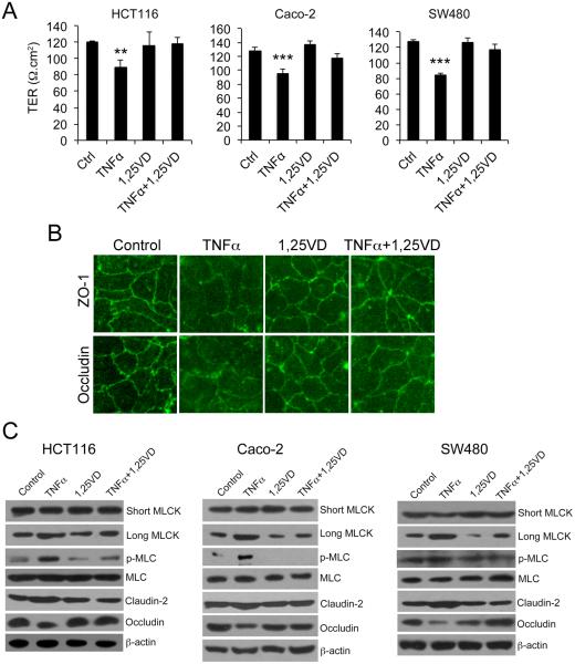
Background: The myosin light chain kinase (MLCK) pathway controls intestinal epithelial barrier permeability by regulating the tight junction. 1,25-dihydroxyvitamin D (1,25(OH)2D3)-vitamin D receptor (VDR) signaling protects the epithelial barrier, but the molecular mechanism is incompletely understood.

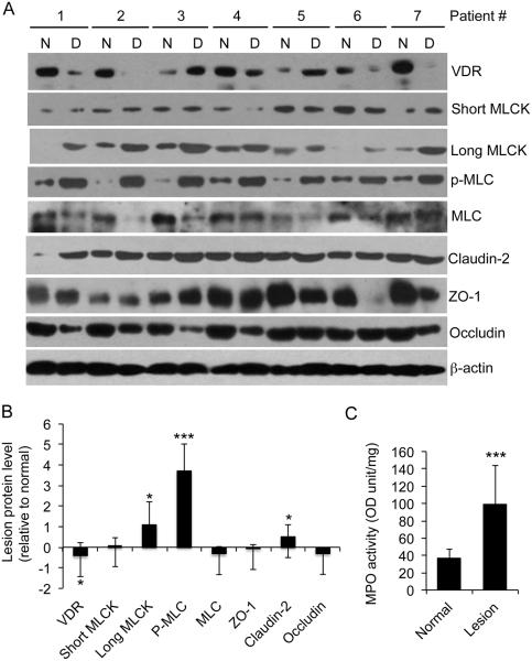
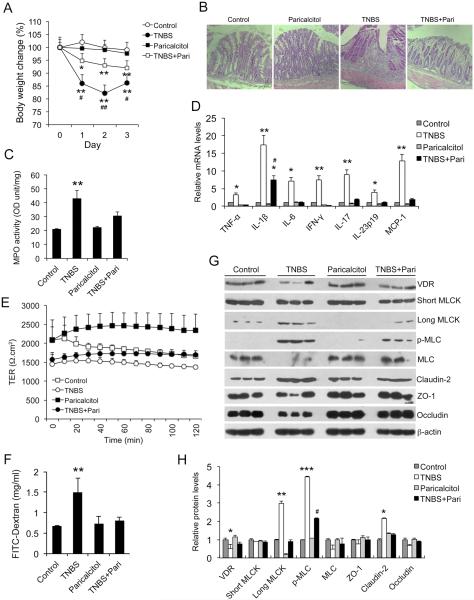
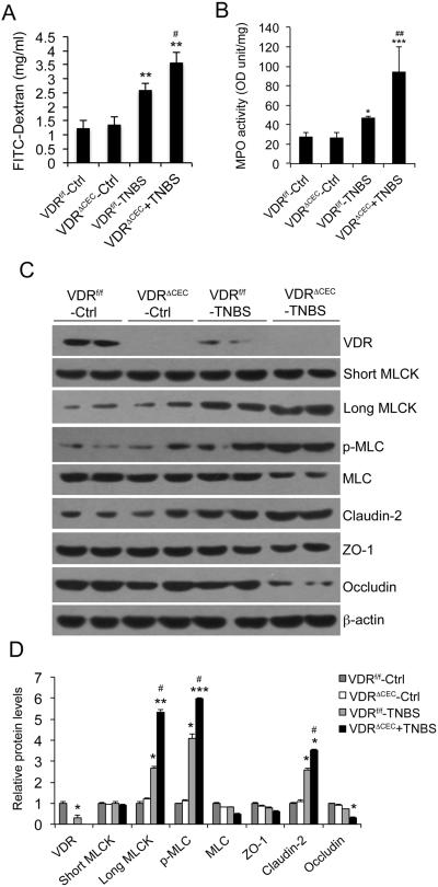
Methods: MLCK activation and barrier permeability were studied using monolayers of HCT116, Caco-2, and SW480 cells treated with tissue necrosis factor α with or without 1,25(OH)2D3. The MLCK pathway was analyzed in normal and inflamed colonic biopsies from patients with ulcerative colitis. Colonic mucosal barrier permeability and MLCK activation were also investigated using trinitrobenzene sulfonic acid-induced colitis models in vitamin D analog paricalcitol-treated wild-type mice and mice carrying VDR deletion in colonic epithelial cells.

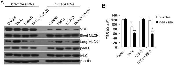
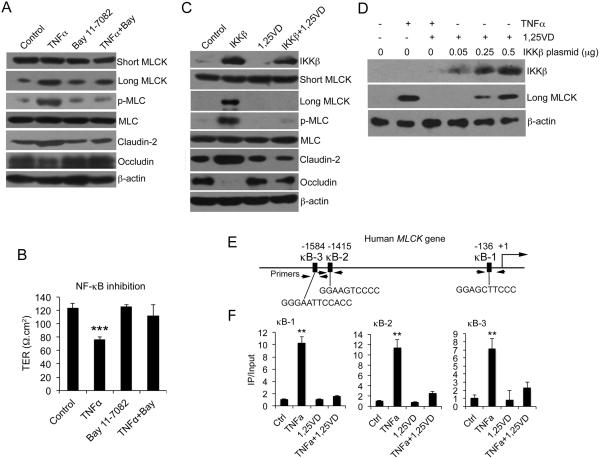
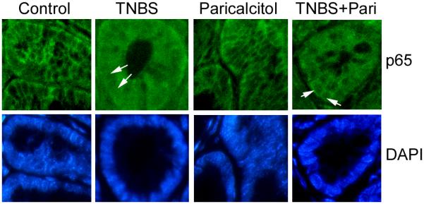
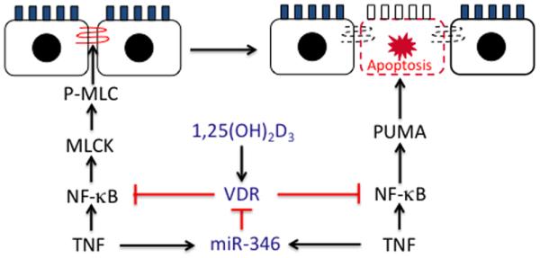
Results: Tissue necrosis factor α increased cell monolayer permeability and induced long isoform of MLCK expression and myosin II regulatory light chain (MLC) phosphorylation, and 1,25(OH)2D3 blocked tissue necrosis factor α-induced increases in monolayer permeability and MLCK-MLC pathway activation by a VDR-dependent fashion. 1,25(OH)2D3 directly suppressed long MLCK expression by attenuating NF-κB activation, and chromatin immunoprecipitation assays confirmed that 1,25(OH)2D3 disrupted p65 binding to 3 κB sites in long MLCK gene promoter. In human ulcerative colitis biopsies, VDR reduction was associated with increases in long MLCK expression and MLC phosphorylation. In trinitrobenzene sulfonic acid colitis models, paricalcitol ameliorated colitis, attenuated the increase in mucosal barrier permeability, and inhibited long MLCK induction and MLC phosphorylation. In contrast, mice with colonic epithelial VDR deletion exhibited more robust increases in mucosal barrier permeability and MLCK activation compared with wild-type mice.

Conclusions: These data demonstrate that 1,25(OH)2D3-VDR signaling preserves the mucosal barrier integrity by abrogating MLCK-dependent tight junction dysregulation during colonic inflammation.


Figures

Figure 1
Figure 1
1,25(OH)2D3 counteracts the effects of TNF-α on paracellular permeability and MLCK activation. (A) Transepithelial resistance (TER) of monolayers formed by HCT-116, Caco-2 and SW480 cells under different treatment as indicated. The cells were grown on transwells and treated or untreated with TNF-α±1,25(OH)2D3 for 12 hours. (B) Caco-2 monolayer immunostained with antibodies against ZO-1 or occludin as indicated. Magnification, 200x. (C) Western blot analyses of HCT116, Caco-2 and SW480 cells under different treatments as indicated. The blots were analyzed with a number of antibodies against short MLCK, long MLCK, phospho-MLC, MLC, claudin-2, occludin and β-actin as shown. TER, transepithelial resistance; 1,25-VD, 1,25(OH)2D3; Ctrl, control. ** P<0.01, *** P<0.001 vs. the rest.
Figure 2
Figure 2
1,25(OH)2D3 regulation of MLCK activation and paracellular permeability is dependent on VDR. (A) Western blot analyses of HCT116 cells transfected with scramble or hVDR-specific siRNA followed by various treatments as indicated. The blots were incubated with a number of antibodies as shown. (B) Transepithelial resistance (TER) of HCT116 monolayers transfected with scramble or hVDR-specific siRNA under different treatments as indicated. *P<0.05; ** P<0.01 vs. corresponding Ctrl or 1,25-VD; # P<0.05 vs. corresponding scramble.
Figure 3
Figure 3
1,25(OH)2D3 blocks MLCK activation by targeting NF-κB activation. (A) Western blot analyses of HCT-116 cells treated or untreated with TNF-α ± BAY 11-7082 as indicated. (B) Transepithelial resistance (TER) of HCT116 cell monolayers under different treatments as indicated for 6 hours. *** P<0.001 vs. the rest. (C) HCT116 cells were transfected with empty vector or IKKβ-expressing plasmid, followed by 1,25(OH)2D3 or vehicle treatment as indicated. Cell lysates were analyzed by Western blotting. (D) HCT116 cells were transfected with increasing amount of IKKβ and then treated with 1,25(OH)2D3 as shown. Cell lysates were analyzed by Western blotting. (E) Schematic illustration of human MLCK gene promoter containing three putative κB sites (κB-1, κB-2 and κB-3) at −136, −1451 and −1584 as indicated. The PCR primer locations were also indicated. (F) ChIP assays using anti-p65 antibodies for HCT116 cells pre-treated with 1,25(OH)2D3 or vehicle followed by 4-hour TNF-α or saline treatment as indicated. The results for κB-1, κB-2 and κB-3 sites are shown. Similar results were seen for 10-hour treatment. **P<0.01 vs. the rest.
Figure 4
Figure 4
Reduction in VDR expression is associated with MLCK pathway activation in colonic biopsies from in ulcerative colitis patients. (A) Western blot analyses of inflamed lesion biopsies and adjacent normal tissues from seven ulcerative colitis patients using antibodies as indicated. (B) Relative protein levels in the lesions compared to the normal tissues, determined by densitometric quantitation of protein bands on the blots. (C) MPO activity in the normal and lesion biopsies. *P<0.05; ** P<0.01; ***P<0.001 vs. normal; n=7. N, normal adjacent tissue; D, diseased tissue.
Figure 5
Figure 5
Paricalcitol preserves mucosal barrier permeability, attenuates MLCK activation and ameliorates colitis in TNBS colitis model. (A) Mouse body weight changes in four treatment groups following TNBS instillation. *P<0.05, **P<0.01 vs. Ctrl and Paricalcitol; #P<0.05 vs. TNBS+Pari; n=5 in each group. (B) H&E staining of distal colon sections in four treatment groups of mice on day 3. (C) MPO activity in mucosal lysates from four groups of mice on day 3. **P<0.01 vs. the rest; n=5 in each group. (D) Real time RT-PCR quantitation of mucosal pro-inflammatory cytokines and chemokines in the four groups of mice on day 3. *P<0.05; ** P<0.01 vs. the rest; #P<0.05 vs. TNBS; n=5 in each genotype. (E) Transepithelial resistance (TER) of colon mucosa from control mice and mice treated with paricalcitol, TNBS and TNBS+Paricalcitol on day 1. (F) Quantitation of paracellular FITC-dextran passage across mucosal barrier in mice from the four treatment groups. ** P<0.01 vs. the rest. (G and H) Western blot analyses (G) and densitometric quantitation (H) of colonic mucosal lysates from the four treatment groups. *P<0.05, **P<0.01, ***P<0.001 vs. the rest for the same protein; #P<0.05 vs. TNBS; n=3.
Figure 6
Figure 6
Immunostaining of colon sections from control mice and mice treated with paricalcitol, TNBS and TNBS+Paricalcitol with antibody against NF-κB p65. The nuclei were stained with DAPI. Arrows indicate nuclei. Note that p65 is translocated in TNBS-treated colonocytes, and the p65 nuclear translocation is markedly attenuated by paricalcitol in colonocytes treated with TNBS+pariocalcitol.
Figure 7
Figure 7
Epithelial hVDR deletion exacerbates MLCK activation and increases mucosal permeability in TNBS colitis model. (A) Paracellular FITC-dextran absorption across the barrier in the four groups of mice. **P<0.01 vs. two Ctrls; #P<0.05 vs. WT+TNBS. (B) Mucosal MPO activity. **P<0.01 vs. two Ctrls; #P<0.05 vs. WT+TNBS. (C) Western blot analyses of mucosal lysates in the four groups of mice on day 2. (D) Densitometric quantitation of Western blot data. *P<0.05, ** P<0.01; *** P<0.001 vs. two Ctrls; #P<0.05 vs. WT+TNBS; n=5 in each group.
Figure 8
Figure 8
1,25(OH)2D3-VDR signaling protects the mucosal barrier. In early stage of colitis development, 1,25(OH)2D3-VDR signaling preserves tight junction barrier by down-regulating long MLCK (this study). With colitis progress, 1,25(OH)2D3-VDR inhibits MLCK-independent epithelial cell apoptosis by down-regulating PUMA (see ref. 25). The mechanism of both regulatory pathways is the blockade of NF-κB activation. Conversely, TNF-α, via induction of miR-346, down-regulates epithelial VDR to attenuate its protective effects (see ref. 26).

References

    1. Laukoetter MG, Bruewer M, Nusrat A. Regulation of the intestinal epithelial barrier by the apical junctional complex. Curr Opin Gastroenterol. 2006;22:85–89. - PubMed
    1. Gibson PR. Increased gut permeability in Crohn's disease: is TNF the link? Gut. 2004;53:1724–1725. - PMC - PubMed
    1. Fasano A, Shea-Donohue T. Mechanisms of disease: the role of intestinal barrier function in the pathogenesis of gastrointestinal autoimmune diseases. Nat Clin Pract Gastroenterol Hepatol. 2005;2:416–422. - PubMed
    1. Su L, Nalle SC, Shen L, et al. TNFR2 activates MLCK-dependent tight junction dysregulation to cause apoptosis-mediated barrier loss and experimental colitis. Gastroenterology. 2013;145:407–415. - PMC - PubMed
    1. Cunningham KE, Turner JR. Myosin light chain kinase: pulling the strings of epithelial tight junction function. Ann N Y Acad Sci. 2012;1258:34–42. - PMC - PubMed

Publication types

MeSH terms