. 2015 Dec;135(12):3133-3143.
doi: 10.1038/jid.2015.314. Epub 2015 Aug 19.

Increased Susceptibility to Skin Carcinogenesis Associated with a Spontaneous Mouse Mutation in the Palmitoyl Transferase Zdhhc13 Gene

Affiliations

Increased Susceptibility to Skin Carcinogenesis Associated with a Spontaneous Mouse Mutation in the Palmitoyl Transferase Zdhhc13 Gene

Carlos J Perez et al. J Invest Dermatol. 2015 Dec.

Abstract

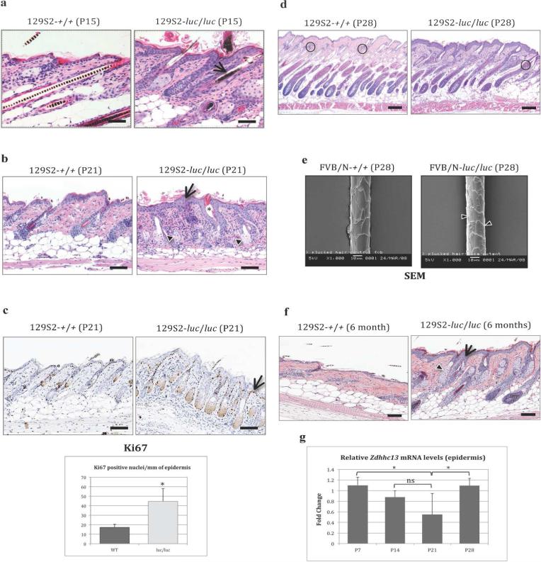
Here we describe a spontaneous mutation in the Zdhhc13 (zinc finger, DHHC domain containing 13) gene (also called Hip14l), one of 24 genes encoding palmitoyl acyltransferase (PAT) enzymes in the mouse. This mutation (Zdhhc13luc) was identified as a nonsense base substitution, which results in a premature stop codon that generates a truncated form of the ZDHHC13 protein, representing a potential loss-of-function allele. Homozygous Zdhhc13luc/Zdhhc13luc mice developed generalized hypotrichosis, associated with abnormal hair cycle, epidermal and sebaceous gland hyperplasia, hyperkeratosis, and increased epidermal thickness. Increased keratinocyte proliferation and accelerated transit from basal to more differentiated layers were observed in mutant compared with wild-type (WT) epidermis in untreated skin and after short-term 12-O-tetradecanoyl-phorbol-13-acetate treatment and acute UVB exposure. Interestingly, this epidermal phenotype was associated with constitutive activation of NF-κB (RelA) and increased neutrophil recruitment and elastase activity. Furthermore, tumor multiplicity and malignant progression of papillomas after chemical skin carcinogenesis were significantly higher in mutant mice than WT littermates. To our knowledge, this is the first report of a protective role for PAT in skin carcinogenesis.


Figures

Figure 1
Figure 1. Skin phenotype in Zdhhc13-deficient luc/luc mice
(a) Sparse hair coat, erythema of the muzzle, and redness of the skin in a 129S2/SvPas-luc/luc mouse at P15 (right). Control littermate is shown on the left. (b) P21 FVB/N-luc/luc mouse (left) showing typical generalized alopecia at this stage. P30 FVB/N-luc/luc mouse (right) with characteristic multifocal patchy alopecia affecting the face (particularly in the periocular region) and short vibrissae. In mice older than 12 months, multifocal alopecia and erythema were associated with inflammatory cell infiltrations in the upper follicular lumen (not shown). WT, wild-type.
Figure 2
Figure 2. The luca mutation corresponds to a premature stop codon in the Zdhhc13 gene
(a) The T to A transversion found in exon 7 of the mutant gene generates a premature stop codon (TAA replacing TTA) and a predicted truncated protein. (b) Diagram comparing WT ZDHHC13 with the predicted truncated protein from the mutant allele, lacking 3 out of 6 ankyrin (ANK) repeats, all transmembrane (TM) domains, and the DQHC PAT motif. (c) The graph shows the mean values of fold-change for Zdhhc13 mRNA levels in WT relative to mutant lung. Control and mutant values are relative to those of internal control β-actin. Bar graph values represent mean ± SEM (*p<0.008, one-tailed unpaired Student's t-test). (d) Representative images of in situ hybridization in WT and mutant skin (P15). The transverse sections of the anagen HFs show a highly reduced hybridization signal for Zdhhc13 mRNA in mutant skin relative to WT. (e) Localization of ZDHHC13 protein in NIH3T3 cells. While GFP-ZDHHC13-WT protein (green) localizes at the Golgi apparatus (white box), mutant GFP-ZDHHC13-truncated protein is localized throughout the cytoplasm and nucleus (red box). (f) Protein palmitoylation in WT and mutant skin. Data are shown as mean ± SEM (a,b: significant with p-value <0.01). Bars = 100 μm (d). DAPI, 4',6-Diamidino-2-phenylindole; GFP, green fluorescent protein; HA, hydroxylamine; HF, hair follicle; WT, wild type.
Figure 3
Figure 3. Histology of the 129S2/SvPas-Zdhhc13luc/Zdhhc13luc mutant skin
(a) H&E-stained dorsal skin sections from untreated WT and mutant at P15. Mutant skin shows mild epidermal hyperplasia with hyperkeratosis. Arrow shows pigment clumps in a mutant. (b) H&E-stained skin sections from WT and mutant at P21. Mutant skin shows markedly thickened epidermis (arrow) with orthokeratotic hyperkeratosis, HF dysplasia, and SG hyperplasia. Arrowheads show SG hyperplasia. Asterisk indicates dilated infundibulum. (c) Ki67-stained sections from WT and mutant at P21. Mutant skin exhibits increased number of positive nuclei in the basal and suprabasal layers of the epidermis (arrow). The bar graph represents the mean value ± SD, n=4 (*p<0.03, independent two samples t-test). (d) H&E sections at P28 showing anagen-stage HFs. Mutant skin shows slight delay in HF cycling, fewer old club hairs (circles), and epidermal and SG hyperplasia. (e) SEM image of WT HS (left) demonstrating normal arrangement of distally-oriented overlapping cuticular scales. Right, mutant HS demonstrating abnormal cuticular scales with rough surface and ill-defined borders (arrowheads). (f) Mutant skin at 6 months showing pronounced epidermal (arrow) and SG hyperplasia (arrowhead). (g) The graph shows the mean values of fold-change for Zdhhc13 mRNA levels in WT epidermis at P7, P14, P21, and P28. Control values are relative to those of internal control (mouse GAPDH). Bar graph values represent mean ± SEM (*p<0.08, ns: no significance, one tailed unpaired Student's t-test). Bars = 40 μm (a) and 100 μm (b, d and f). SEM bar = 10 μm (e). H&E, hematoxylin and eosin; HF, hair follicle; HS, hair shaft; WT, wild-type.
Figure 4
Figure 4. Increased keratinocyte proliferation and transit in mutant epidermis after acute TPA and UVB exposure
(a) Bar graphs show epidermal thickness in μm (left) and cell proliferation level as BrdU-positive nuclei per mm of epidermis (right) after TPA. Values represent mean ± SD (*p<0.01 both graphs). (b) Representative BrdU-immunostaining of TPA-treated dorsal skin from WT and mutant after 8 h of BrdU. Progression of BrdU-labeled keratinocytes towards the surface is faster in mutant epidermis (arrows show positively stained nuclei in granular layer of mutant skin, not seen in WT). (c) Representative BrdU-immunostaining of untreated dorsal skin from WT and mutant after BrdU (arrows show increased number of positive nuclei in the basal layer of mutant epidermis). Bar graph represents the mean value ± SD, n=4 (*p<0.008, independent two-samples t-test). (d-g) Representative IHC images showing localization of Ki67-, p-STAT3-, p21-, and p53-positive keratinocytes in UVB-exposed skin from WT and mutant. Arrows indicate strong positively stained nuclei in all layers of the epidermis in mutant skin, not seen in WT. Bar graphs represent mean value ± SD, n=4 (*p<0.003 for Ki67 and p-STAT3; *p<0.001 for p21 and p53, independent two-samples t-test). Bars = 40 μm (b, d, f and g) and 100 μm (c and e). BrdU, 5-bromo-2-deoxyuridine; IHC, immunohistochemistry; SD, standard deviation; TPA, 12-O-tetradecanoylphorbol-13-acetate; WT, wild-type.
Figure 5
Figure 5. Constitutive activation of NF-κB and increased neutrophil recruitment in the mutant skin
(a, b) Representative IHC showing NF-κB expression in untreated dorsal skin. Strong positively stained nuclei (over-phosphorylated p65) are present in all layers of mutant epidermis (arrows), ORS (big arrowheads), and SGs (small arrowheads), not seen in WT. Bar graph: mean value ± SD, n=4 (*p<0.001, independent two-samples t-test). (c) Over-phosphorylation of p65 in mutant skin after 4 TPA treatments. Arrows show positively-stained keratinocytes on the granular layer of mutant skin 8 h after last TPA. Arrowheads indicate positively-stained nuclei in ORS. Bar graph: mean value ± SD, n=4 (*p<0.001, independent two samples t-test). (d) WB from total and phospho(Ser536)-NF-κB protein levels in untreated skin. Mean ± SEM, n=3 (*p<0.01, Student's t-test). (e) Whole-mouse fluorescence images of neutrophil elastase activity after UV and TPA. Color scale indicates radiant efficiency. Quantitative analysis of fluorescence levels with mutant mice showing significantly stronger signal than WT (UV-*p<0.007; TPA-*p<0.001, n=4). (f) H&E- and LY6G-stained sections from UVB-treated skin. Mutant skin shows markedly thickened epidermis, increased inflammatory infiltration and higher levels of LY6G-positive cells (neutrophils) in the dermis (arrows). (g) Similar results as in (f) in TPA-treated skin. Bars = 100 μm (a, c) and 40 μm (b, f, g). WT, wild-type; TPA, 12-O-tetradecanoylphorbol-13-acetate; H&E, hematoxylin and eosin.
Figure 6
Figure 6. Increased susceptibility to skin carcinogenesis in Zdhhc13luc/Zdhhc13luc mutant mice
(a) Multiplicity of skin tumors after two stage carcinogenesis in FVB/N-luc/luc mice (n = 10) was highly statistically significant compared to that of WT littermates (n = 10) at 15 wk of TPA treatment (*p<0.001, Wilcoxon Rank Sum test). The number of papillomas was determined weekly. Tumor multiplicity is the average number of skin tumors per mouse. (b) Representative images of DMBA/TPA-induced skin tumors after 15 wk of TPA in WT (left) and mutant (right) FVB/N mice. (c-f) Representative IHC staining for E-cadherin, MMP9, Slug, and Snail in DMBA/TPA induced skin papillomas from WT and mutant mice. The papillomas from mutant mice show reduced levels of E-cadherin and higher expression of Slug, Snail, and MMP9 when compared with WT (arrows). This is in agreement with the increased invasiveness observed in mutant tumors. Bar = 200 μm. WT, wild type; wk, weeks; TPA, 12-O-tetradecanoylphorbol-13-acetate; DMBA, 7, 12-dimethylbenz [a] anthracene.

References

    1. Benavides F, Blando J, Contreras O, et al. Protective role of cathepsin L in mouse skin carcinogenesis. Mol Carcinog advance online publication. 2011 May 2; DOI. - PMC - PubMed
    1. Benavides F, Starost MF, Flores M, et al. Impaired hair follicle morphogenesis and cycling with abnormal epidermal differentiation in nackt mice, a cathepsin L-deficient mutation. Am J Pathol. 2002;161:693–703. - PMC - PubMed
    1. Benavides F, Stern MC, Glasscock E, et al. Microsatellite DNA variants between the inbred SENCAR mouse strains. Mol Carcinog. 2000;28:191–5. - PubMed
    1. Berger MF, Hodis E, Heffernan TP, et al. Melanoma genome sequencing reveals frequent PREX2 mutations. Nature. 2012;485:502–6. - PMC - PubMed
    1. Budunova IV, Perez P, Vaden VR, et al. Increased expression of p50-NF-kappaB and constitutive activation of NF-kappaB transcription factors during mouse skin carcinogenesis. Oncogene. 1999;18:7423–31. - PubMed

Publication types