. 2015 Jul-Sep;25(3):213-25.
doi: 10.4103/0971-3026.161431.

Chest tuberculosis: Radiological review and imaging recommendations

Affiliations

Chest tuberculosis: Radiological review and imaging recommendations

Ashu Seith Bhalla et al. Indian J Radiol Imaging. 2015 Jul-Sep.

Abstract

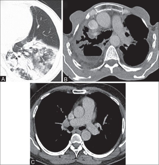
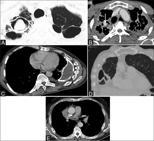
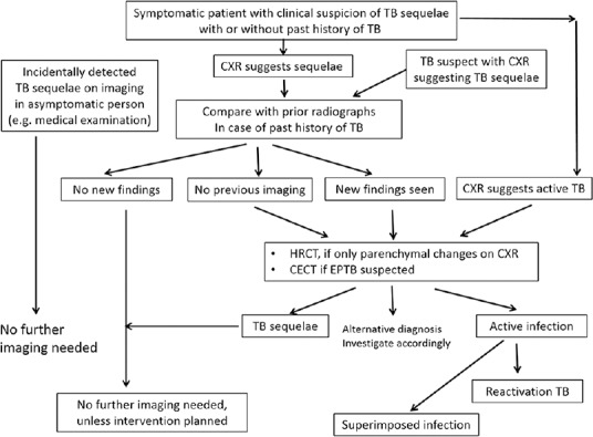
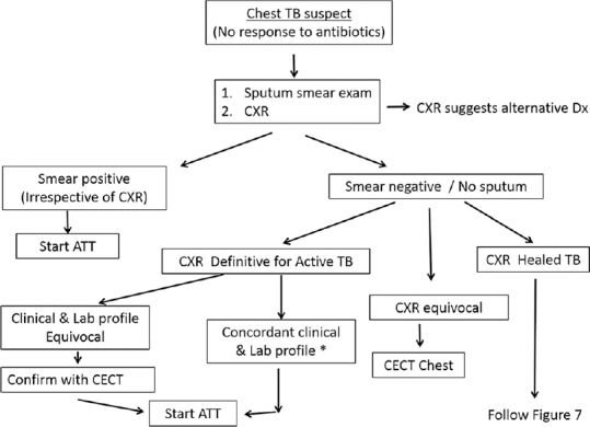
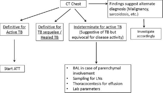
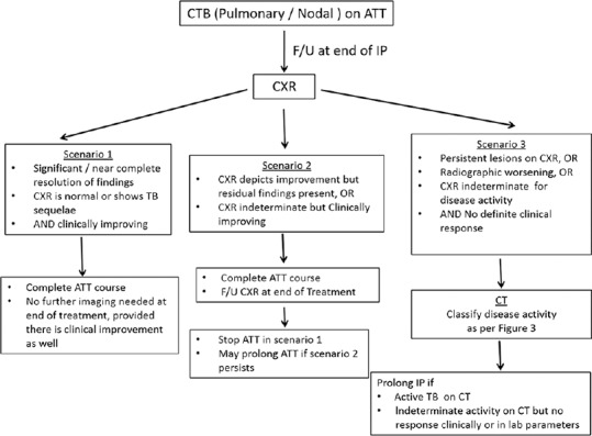
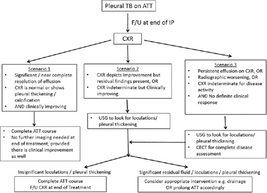
Chest tuberculosis (CTB) is a widespread problem, especially in our country where it is one of the leading causes of mortality. The article reviews the imaging findings in CTB on various modalities. We also attempt to categorize the findings into those definitive for active TB, indeterminate for disease activity, and those indicating healed TB. Though various radiological modalities are widely used in evaluation of such patients, no imaging guidelines exist for the use of these modalities in diagnosis and follow-up. Consequently, imaging is not optimally utilized and patients are often unnecessarily subjected to repeated CT examinations, which is undesirable. Based on the available literature and our experience, we propose certain recommendations delineating the role of imaging in the diagnosis and follow-up of such patients. The authors recognize that this is an evolving field and there may be future revisions depending on emergence of new evidence.

Keywords: Chest radiograph; TB active; chest tuberculosis (pulmonary; computed tomography; imaging recommendations; nodal and pleural).


Conflict of interest statement

Conflict of Interest: None declared.

Figures

Figure 1
Figure 1
Natural history of chest tuberculosis
Figure 2 (A-E)
Figure 2 (A-E)
Chest radiographs in active TB. (A) CXR depicts RT upper zone consolidation with prominent RT hilum. (B) CXR in a different patient shows multiple coalescent air-space nodules in RT upper zone. (C) CXR in a different patient shows multiple ill-defined reticulo-nodular lesions in both lungs with basal predominance, suggestive of miliary TB. (D) CXR in a different patient shows active post-primary TB. Cavity with surrounding consolidation is seen in LT upper zone. Scattered air-space nodules are seen in both lungs with left hilar adenopathy. (E) There is RT-sided loculated pleural effusion with multiple air-space nodules scattered in both lungs
Figure 3 (A-G)
Figure 3 (A-G)
Chest CT in active TB. (A) Lung window (window center -600 HU, width 1200 HU) of chest CT depicts centrilobular and clustered nodules in posterior segment of RT upper lobe, suggesting active endobronchial infection. (B) Lung window of chest CT in a different patient shows cavity with surrounding consolidation in apicoposterior segment of LT upper lobe. Multiple centrilobular nodules with tree-in-bud branching pattern are also seen. (C) Sagittal multiplanar CT reformat lung window shows segmental consolidations in RT upper lobe. (D) Axial CT section lung window (in the same patient as in Figure 1c) in miliary TB shows multiple tiny discrete nodules randomly distributed in both lungs. (E) Axial CECT mediastinal window (window center 40 HU, width 400 HU) shows conglomerate rim-enhancing lymphadenopathy in paratracheal locations and multiple enlarged lymph nodes in prevascular location as well showing central necrosis. (F) Axial CECT mediastinal window shows RT-sided effusion with enhancing layers of pleura (split pleura sign) and rib crowding suggestive of empyema. (G) Axial non-contrast CT mediastinal window shows LT-sided free effusion
Figure 4(A-F)
Figure 4(A-F)
Imaging features of healed TB. (A) CXR shows thin-walled cavity in left upper zone. Areas of fibro-bronchiectasis and fibrocalcific lesions are seen in left upper zone, RT upper and mid zones. (B) Axial CT lung window (window center -600 HU, width 1200 HU) shows clustered thin-walled cavities in superior segment RT lower lobe. (C) CXR shows volume loss in both upper zones with apical pleural thickening, pulled hila, fibro-bronchiectasis, and calcific foci. (D) CXR shows fibro-bronchiectasis both upper zones. (E) CECT mediastinal window (window center 40 HU, width 400 HU) shows left-sided pleural thickening and focal plaque-like calcifications. (F) CT lung window section in end-stage lung disease shows collapse and bronchiectasis involving the left lung with ipsilateral mediastinal shift and rib crowding
Figure 5 (A-C)
Figure 5 (A-C)
Imaging features indeterminate for disease activity in CTB. (A) Axial CT lung window (window center -600 HU, width 1200 HU) shows consolidation in basal segments of left lower lobe. No ipsilateral adenopathy, no cavitation was seen. (B) CECT mediastinal window (window center 40 HU, width 400 HU) shows cavity with air-fluid level in RT upper lobe. Note is also made of bronchiectasis and apical pleural thickening. (C) Axial CECT mediastinal window shows small discrete homogeneous lymph node in RT hilar location, measuring ~10 mm in short axis dimension (SAD). This node was unchanged in size and morphology after complete course of ATT
Figure 6(A-E)
Figure 6(A-E)
Imaging findings in tuberculous complications. (A) Axial CT lung window (window center -600 HU, width 1200 HU) shows thin-walled cavities in both upper lobes and presence of aspergilloma in RT upper lobe cavity. (B) Axial CECT mediastinal window (window center 40 HU, width 400 HU) shows contrast-filled pseudoaneurym (arrow) arising from the superior division of RT pulmonary artery (Rasmussen aneurysm) in the background of fibro-cavitary lesions in both upper lobes. (C) Axial CECT mediastinal window shows chronic empyema LT side with volume loss and pleural calcifications. (D) Coronal CT lung window depicts abnormal communication of pleural space with bronchial tree suggesting a bronchopleural fistula. (E) Axial CECT mediastinal window shows calcified LN in RT hilum causing post-obstructive atelectasis of RT middle lobe
Figure 7
Figure 7
Imaging work-up of symptomatic patients suspected/detected to have TB sequelae
Figure 8
Figure 8
Flowchart depicting the role of imaging in diagnosis of CTB (*lab profile includes hematological parameters like ESR, CRP, TLC, DLC, and Mantoux test)
Figure 9
Figure 9
Flowchart demonstrating the stratification of patients after CT. Based on CT findings, the patient should be categorized into either active TB, healed TB, or indeterminate for disease activity
Figure 10
Figure 10
Imaging protocol for follow-up of PTB and mediastinal nodal TB (IP = Intensive phase)
Figure 11
Figure 11
Imaging protocol for follow-up of pleural TB (IP = Intensive phase)

References

    1. Foulds J, O’Brien R. New tools for the diagnosis of tuberculosis: The perspective of developing countries. Int J Tuberc Lung Dis. 1998;2:778–83. - PubMed
    1. Okur E, Yilmaz A, Saygi A, Selvi A, Süngün F, Oztürk E, et al. Patterns of delays in diagnosis amongst patients with smear-positive pulmonary tuberculosis at a teaching hospital in Turkey. Clin Microbiol Infect. 2006;12:90–2. - PubMed
    1. Im JG, Itoh H, Shim YS, Lee JH, Ahn J, Han MC, et al. Pulmonary tuberculosis: CT findings- early active disease and sequential change with antituberculous therapy. Radiology. 1993;186:653–60. - PubMed
    1. Jeong YJ, Lee KS. Pulmonary tuberculosis: Up-to-date imaging and management. AJR Am J Roentgenol. 2008;191:834–44. - PubMed
    1. WHO Global tuberculosis report. 2013. [Last accessed on 2014 Aug 06]. Available from: http://www.who.int/tb .