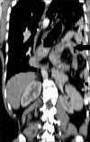
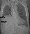
Blunt traumatic diaphragmatic hernia: Pictorial review of CT signs
- PMID: 26288515
- PMCID: PMC4531445
- DOI: 10.4103/0971-3026.161433
Blunt traumatic diaphragmatic hernia: Pictorial review of CT signs
Abstract
Blunt diaphragmatic rupture rarely accounts for immediate mortality and may go clinically silent until complications occur which can be life threatening. Although many imaging techniques have proven useful for the diagnosis of blunt diaphragmatic rupture, multidetector CT (MDCT) is considered to be the reference standard for the diagnosis of diaphragmatic injury. Numerous CT signs indicating blunt diaphragmatic rupture have been described in literature with variable significance. Accurate diagnosis depends upon the analysis of all the signs rather than a single sign; however, the presence of blunt diaphragmatic rupture should be considered in the presence of any of the described signs. We present a pictorial review of various CT signs used to diagnose blunt diaphragmatic injury. Multiplanar reconstruction is very useful; however, predominantly axial sections have been described in this pictorial review as the images shown are from dual-slice CT.
Keywords: Computed tomography; diaphragm; hernia; injury; trauma.
Conflict of interest statement
Figures


Similar articles
-
The dangling diaphragm sign: sensitivity and comparison with existing CT signs of blunt traumatic diaphragmatic rupture.Emerg Radiol. 2010 Jan;17(1):37-44. doi: 10.1007/s10140-009-0819-5. Epub 2009 May 16. Emerg Radiol. 2010. PMID: 19449046
-
The Role of Computed Tomography in the Diagnostics of Diaphragmatic Injury After Blunt Thoraco-Abdominal Trauma.Pol J Radiol. 2016 Nov 4;81:522-528. doi: 10.12659/PJR.897866. eCollection 2016. Pol J Radiol. 2016. PMID: 27867441 Free PMC article.
-
CT of blunt diaphragmatic rupture.Radiographics. 2012 Mar-Apr;32(2):477-98. doi: 10.1148/rg.322115082. Radiographics. 2012. PMID: 22411944 Review.
-
Computed Tomography in the Evaluation of Diaphragmatic Hernia following Blunt Trauma.Indian J Surg. 2012 Aug;74(4):288-93. doi: 10.1007/s12262-011-0390-7. Epub 2012 Jan 10. Indian J Surg. 2012. PMID: 23904715 Free PMC article.
-
Imaging of diaphragmatic rupture after trauma.Clin Radiol. 2006 Jun;61(6):467-77. doi: 10.1016/j.crad.2006.02.006. Clin Radiol. 2006. PMID: 16713417 Review.
Cited by
-
Fractured Ribs and the CT Funky Fat Sign of Diaphragmatic Rupture.Case Rep Radiol. 2016;2016:6723632. doi: 10.1155/2016/6723632. Epub 2016 Jun 26. Case Rep Radiol. 2016. PMID: 27429823 Free PMC article.
-
Bilateral delayed traumatic diaphragmatic injury.J Surg Case Rep. 2021 Apr 14;2021(4):rjab052. doi: 10.1093/jscr/rjab052. eCollection 2021 Apr. J Surg Case Rep. 2021. PMID: 33884164 Free PMC article.
-
Giant Traumatic Diaphragmatic Hernia: A Report of Delayed Presentation.Cureus. 2021 Dec 10;13(12):e20315. doi: 10.7759/cureus.20315. eCollection 2021 Dec. Cureus. 2021. Retraction in: Cureus. 2024 Jan 25;16(1):r111. doi: 10.7759/cureus.r111. PMID: 35028214 Free PMC article. Retracted.
-
Laparoscopic Repair of Blunt Traumatic Diaphragmatic Hernia.Cureus. 2023 Sep 26;15(9):e46017. doi: 10.7759/cureus.46017. eCollection 2023 Sep. Cureus. 2023. PMID: 37900497 Free PMC article.
-
A delayed presentation of traumatic diaphragmatic hernia in a young male: a unusual case report and comprehensive review of literature.J Surg Case Rep. 2024 Oct 2;2024(10):rjae613. doi: 10.1093/jscr/rjae613. eCollection 2024 Oct. J Surg Case Rep. 2024. PMID: 39364427 Free PMC article.
References
-
- Nchimi A, Szapiro D, Ghaye B, Willems V, Khamis J, Haquet L, et al. Helical CT of blunt diaphragmatic rupture. AJR Am J Roentgenol. 2005;184:24–30. - PubMed
-
- Chen HW, Wong YC, Wang LJ, Fu CJ, Fang JF, Lin BC. Computed tomography in left-sided and right-sided blunt diaphragmatic rupture: Experience with 43 patients. Clin Radiol. 2010;65:206–12. - PubMed
-
- Murray JG, Caoili E, Gruden JF, Evans SJ, Halvorsen RA, Jr, Mackersie RC. Acute rupture of the diaphragm due to blunt trauma: Diagnostic sensitivity and specificity of CT. AJR Am J Roentgenol. 1996;166:1035–9. - PubMed
-
- Killeen KL, Mirvis SE, Shanmuganathan K. Helical CT of diaphragmatic rupture caused by blunt trauma. AJR Am J Roentgenol. 1999;173:1611–6. - PubMed
LinkOut - more resources
Full Text Sources
Other Literature Sources

