. 2015 Jul-Sep;25(3):246-60.
doi: 10.4103/0971-3026.161445.

Perspectives in ultrasound-guided musculoskeletal interventions

Affiliations

Perspectives in ultrasound-guided musculoskeletal interventions

Aditya Ravindra Daftary et al. Indian J Radiol Imaging. 2015 Jul-Sep.

Abstract

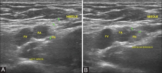
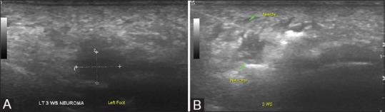
Ultrasonography (USG) is a safe, easily available, and cost-effective modality, which has the additional advantage of being real time for imaging and image-guided interventions of the musculoskeletal system. Musculoskeletal interventions are gaining popularity in sports and rehabilitation for rapid healing of muscle and tendon injuries in professional athletes, healing of chronic tendinopathies, aspiration of joint effusions, periarticular bursae and ganglia, and perineural injections in acute and chronic pain syndromes. This article aims to provide an overview of the spectrum of musculoskeletal interventions that can be done under USG guidance both for diagnostic and therapeutic purposes.

Keywords: Interventions; pain; relief; sports; ultrasound.


Conflict of interest statement

Conflict of Interest: None declared.

Figures

Figure 1 (A and B)
Figure 1 (A and B)
Needle visualization: (A) The needle is best visualized when its long axis is parallel and in line with the long axis of the transducer face and in this plane, the needle is seen as a linear echogenic structure with reverberation artifact distally (B) If the needle is not parallel to the long axis of the transducer as is often seen in deep-seated targets, it may be difficult to identify. Visualization can be improved either by holding the needle steady while moving the transducer to identify the needle or by repositioning the needle to run along the longitudinal axis of the transducer
Figure 2 (A-C)
Figure 2 (A-C)
Tendon sheath injections: (A) Biceps tendon is identified in transverse in the bicipital groove and is traced till its intra-articular portion in the rotator interval, where targeted peritendinous steroid injections are given in cases of tendinosis. Rotator interval is also targeted for injections in adhesive capsulitis which is not controlled by medical therapy alone (B) Peritendinous steroid injections around the peroneal tendon are given as it traverses in the retromalleolar groove or at the site of maximum pain (C) Intratendinous injections with steroid or PRP injections have been attempted in some cases of tendinosis as in the above example of peroneal tendinosis
Figure 3 (A and B)
Figure 3 (A and B)
Peritendinous injections for release of trigger finger: (A) Trigger finger is caused by a tenosynovitis of the digital flexor tendons and presents with snapping fingers. Peritendinous steroid injections provide rapid symptomatic relief by reducing the inflammation. USG guidance, especially in smaller joints, aids and confirms an accurate delivery of the injectate into the area of interest (B) Peritendinous fluid distension confirms delivery of injectate which can be visualized real time with USG
Figure 4 (A-F)
Figure 4 (A-F)
Spectrum of interventions in tennis elbow: (A) In acute tendinosis with minimal underlying tendon abnormality on USG, peritendinous steroids and local anesthetics are used for management, followed by appropriate physiotherapy (B) In cases of acute tendinosis with underlying tears at the common extensor origin, intratendinous steroids or PRP (especially in athletes) have also been attempted accompanied by a graded rehabilitation program (C-F) In chronic calcific tendonitis, a barbotage therapy which involves breakdown of the calcific deposit seen in (C) with continuous needling and irrigation with normal saline (D) till the deposit becomes smaller or more diffuse as seen in (E). (F) Comparison of pre- and post-procedure radiographs of the patient showing a reduction in the size and density of the calcific deposit following the procedure
Figure 5 (A and B)
Figure 5 (A and B)
Acute MCL injuries: (A) Diffusely thickened and altered echotexture of the MCL near its femoral attachment in the left knee with an undersurface hypoechoic area suggesting a tear (single arrow) (B) Intraligamentous PRP injections in acute injuries have been beneficial in athletes, especially during the sporting season wherein rapid recovery and return to activity is desired. Targeted injection of PRP is given at the site of the injury
Figure 6 (A and B)
Figure 6 (A and B)
Painful Pelligrini-Stieda lesion: (A) USG examination shows a calcified focus within the fibers of the MCL referred to as Pelligrini-Stieda lesion (B) Local needling of the calcification followed by steroid injections can be done in occasional cases when these lesions are a cause of unresponsive pain
Figure 7 (A-C)
Figure 7 (A-C)
Subacromial/subdeltoid bursal injections: (A) Subacromial bursa is superficial and easily amenable to USG injections in the management of tendon disorders, subacromial bursitis, and impingement syndromes (B) Needle position within the bursa is confirmed prior to injection (C) Bursal distension during the injection is confirmatory of accurate delivery of injectate
Figure 8(A-C)
Figure 8(A-C)
Aspiration of wrist ganglia: (A) USG examination revealed a ganglion on the dorsal aspect of the wrist overlying the scaphoid and trapezoid bones (scapho-trapezio-trapezoid ganglion) (B) A slightly thicker needle is used since the material within a ganglion is thicker and repeated fenestration of the ganglion may be required till reduction in size is confirmed (C)
Figure 9(A-C)
Figure 9(A-C)
Aspiration of intra-articular ganglia related to the cruciates: (A) Deep-seated ganglia related to the cruciates in the knee are difficult to visualize. Anterior cruciate ligament ganglion is seen as a hypoechoic intra-articular area (G) when scanned from the posterior aspect of the knee joint (B) Needle position can be confirmed within the ganglion and aspiration/fenestration is attempted till reduction in size or alteration in echogenicity is visualized as in (C). This is then followed by a steroid injection
Figure 10 (A-C)
Figure 10 (A-C)
Parameniscal cyst aspiration: (A) Parameniscal cysts are relatively superficially located in the knee joint and can be visualized in the medial and lateral femoral gutters when scanning along the joint lines. The figure demonstrates a medial parameniscal cyst (B) The cyst is easily accessible with USG guidance. Needle position is confirmed by real-time visualization of the needle (C) Post-procedural scan shows near-complete collapse of the cyst
Figure 11
Figure 11
Targeted approach with USG guidance is especially beneficial for deep-seated joints like the hip and shoulder. The image shows accurate needle placement into the glenohumeral joint space for intra-articular injections
Figure 12 (A and B)
Figure 12 (A and B)
Targeted approach with USG guidance is especially beneficial for smaller peripheral joints like in the hands and feet: (A) USG images of the calcaneocuboid joint in the right foot demonstrate osteoarthritic changes with an overhanging cuboid osteophyte (B) An additional advantage of USG being real time is in osteoarthritic joints where the needle can be manipulated under an osteophyte or osseous spur during the procedure itself
Figure 13 (A and B)
Figure 13 (A and B)
Femoral nerve blocks. (A) Femoral nerve is identified just lateral to the femoral vessels in the inguinal region by its classic “salt and pepper” appearance and being superficial, it is amenable to USG interventions (B) Optimal spread of the injectate is confirmed by presence of a hypoechoic halo surrounding the echogenic nerve (halo sign)
Figure 14 (A and B)
Figure 14 (A and B)
Morton's neuroma: (A) USG examination can easily identify a Morton's neuroma as a hypoechoic nodule in the second to third metatarsal web space (B) Post injection, all neuromas displayed increased echogenicity and/or appearance of fluid surrounding it, confirming localization of the therapeutic mixture

Similar articles

Cited by

References

    1. Louis LJ. Musculoskeletal ultrasound intervention: Principles and advances. Radiol Clin North Am. 2008;46:515–33. vi. - PubMed
    1. Stephens MB, Beutler AI, O’Connor FG. Musculoskeletal injections: A review of the evidence. Am Fam Physician. 2008;78:971–6. - PubMed
    1. Casati A, Vinciguerra F, Scarioni M, Cappelleri G, Aldegheri G, Manzoni P, et al. Lidocaine versus ropivacaine for continuous interscalene brachial plexus block after open shoulder surgery. Acta Anaesthesiol Scand. 2003;47:355–60. - PubMed
    1. Scott DB, Lee A, Fagan D, Bowler GM, Bloomfield P, Lundh R. Acute toxicity of ropivacaine compared with that of bupivacaine. Anesth Analg. 1989;69:563–9. - PubMed
    1. Glaser C, Marhofer P, Zimpfer G, Heinz MT, Sitzwohl C, Kapral S, et al. Levobupivacaine versus racemic bupivacaine for spinal anesthesia. Anesth Analg. 2002;94:194–8. - PubMed