

Evaluation of static and dynamic MRI for assessing response of bone sarcomas to preoperative chemotherapy: Correlation with histological necrosis
- PMID: 26288521
- PMCID: PMC4531451
- DOI: 10.4103/0971-3026.161452
Evaluation of static and dynamic MRI for assessing response of bone sarcomas to preoperative chemotherapy: Correlation with histological necrosis
Abstract
Objectives: Preoperative chemotherapy plays a key role in management of bone sarcomas. Postoperative evaluation of histological necrosis has been the gold standard method of assessing response to preoperative chemotherapy. This study was done to evaluate the efficacy of static and dynamic magnetic resonance imaging (MRI) for assessing response preoperatively.
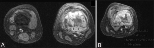
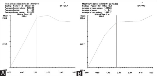
Materials and methods: Our study included 14 patients (12 osteosarcomas and 2 malignant fibrous histiocytomas) with mean age of 21.8 years, treated with preoperative chemotherapy followed by surgery. They were evaluated with static and dynamic MRI twice, before starting chemotherapy and again prior to surgery. Change in tumor volume and slope of signal intensity - time curve were calculated and correlated with percentage of histological necrosis using Pearson correlation test.
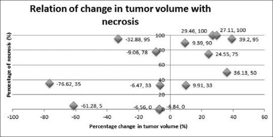
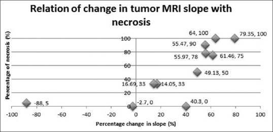
Results: The change in dynamic MRI slope was significant (P = 0.001). Also, ≥60% reduction in slope of the curve proved to be an indicator of good histological response [positive predictive value (PPV) =80%]. Change in tumor volume failed to show significant correlation (P = 0.071). Although it showed high negative predictive value (NPV = 85.7%), PPV was too low (PPV = 57.14%).
Conclusions: Dynamic MRI correctly predicts histological necrosis after administration of preoperative chemotherapy to bone sarcomas. Hence, it can be used as a preoperative indicator of response to neoadjuvant chemotherapy. On the other hand, volumetric assessment by static MRI is not an effective predictor of histological necrosis. This study proves the superiority of dynamic contrast-enhanced study over volumetric study by MRI.
Keywords: Dynamic MR; MR; histological necrosis; malignant fibrous histiocytoma; osteosarcoma; preoperative chemotherapy.
Conflict of interest statement
Figures


References
-
- Glasser DB, Lane JM, Huvos AG, Marcove RC, Rosen G. Survival, prognosis, and therapeutic response in osteogenic sarcoma. The memorial hospital experience. Cancer. 1992;69:698–708. - PubMed
-
- Wunder JS, Paulian G, Huvos AG, Heller G, Meyers PA, Healey JH. The histological response to chemotherapy as a predictor of the oncological outcome of operative treatment of Ewing sarcoma. J Bone Joint Surg Am. 1998;80:1020–33. - PubMed
-
- Kawai A, Sugihara S, Kunisada T, Uchida Y, Inoue H. Imaging assessment of the response of bone tumors to preoperative chemotherapy. Clin Orthop Relat Res. 1997;337:216–25. - PubMed
-
- Raymond AK, Chawla SP, Carrasco CH, Ayala AG, Fanning CV, Grice B, et al. Osteosarcoma chemotherapy effect: A prognostic factor. Semin Diagn Pathol. 1987;4:212–36. - PubMed
-
- Rosen G, Caparros B, Huvos AG, Kosloff C, Nirenberg A, Cacavio A, et al. Preoperative chemotherapy for osteogenic sarcoma: Selection of postoperative adjuvant chemotherapy based on the response of the primary tumor to preoperative chemotherapy. Cancer. 1982;49:1221–30. - PubMed
LinkOut - more resources
Full Text Sources
Other Literature Sources