. 2015 Nov-Dec;17(6):996-1005.
doi: 10.4103/1008-682X.159722.

NODAL secreted by male germ cells regulates the proliferation and function of human Sertoli cells from obstructive azoospermia and nonobstructive azoospermia patients

Affiliations

NODAL secreted by male germ cells regulates the proliferation and function of human Sertoli cells from obstructive azoospermia and nonobstructive azoospermia patients

Ru-Hui Tian et al. Asian J Androl. 2015 Nov-Dec.

Abstract

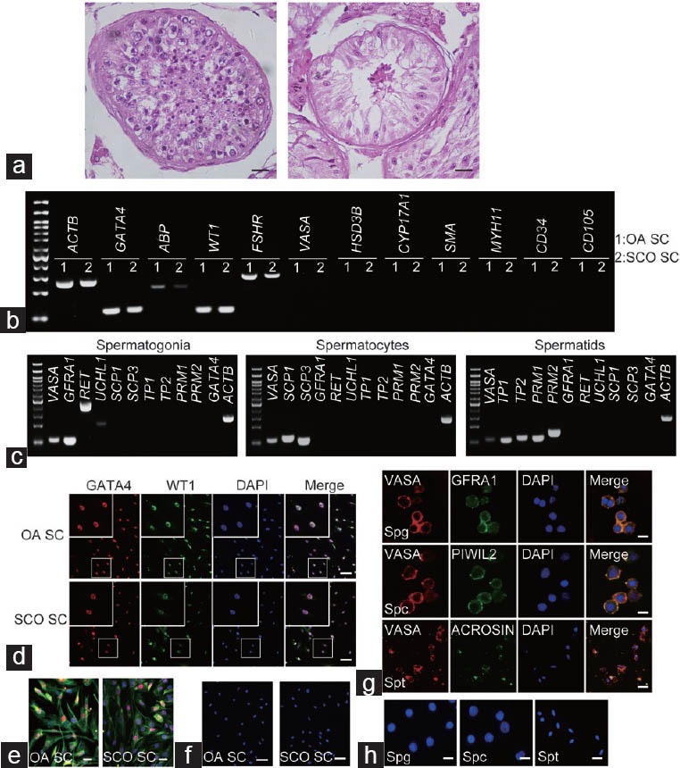
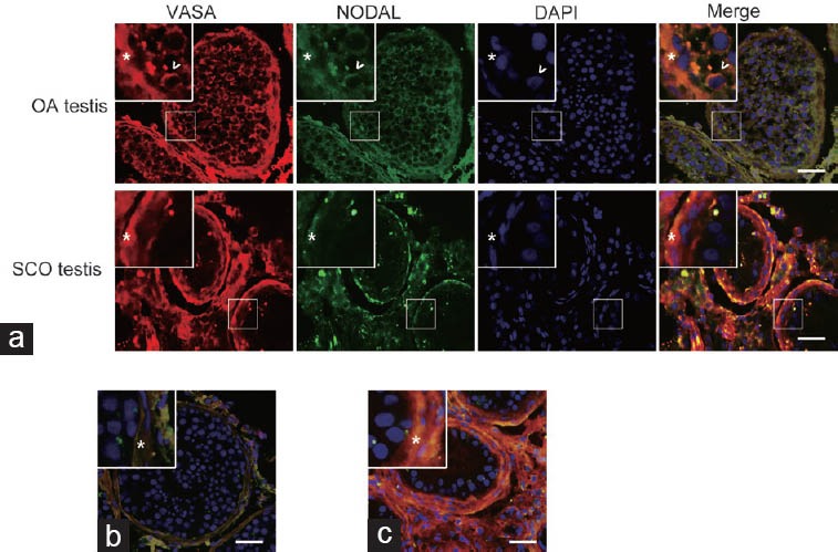
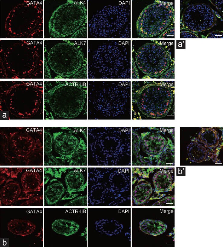
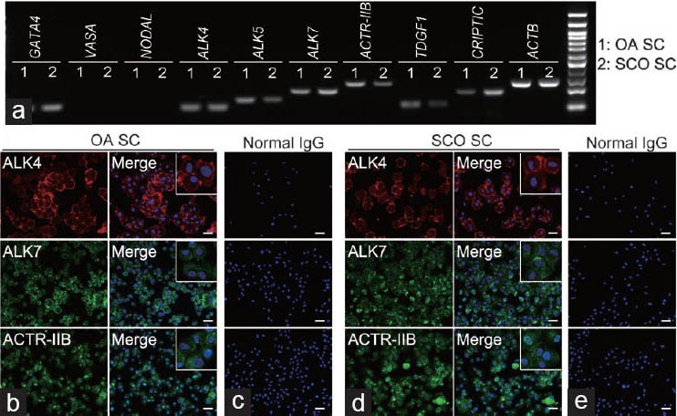
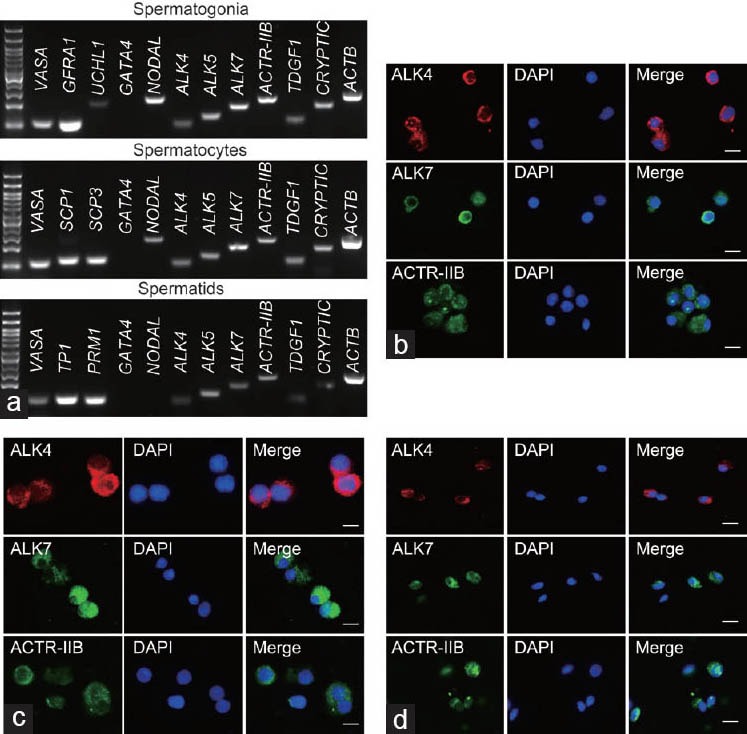
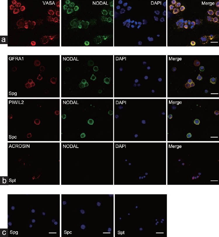
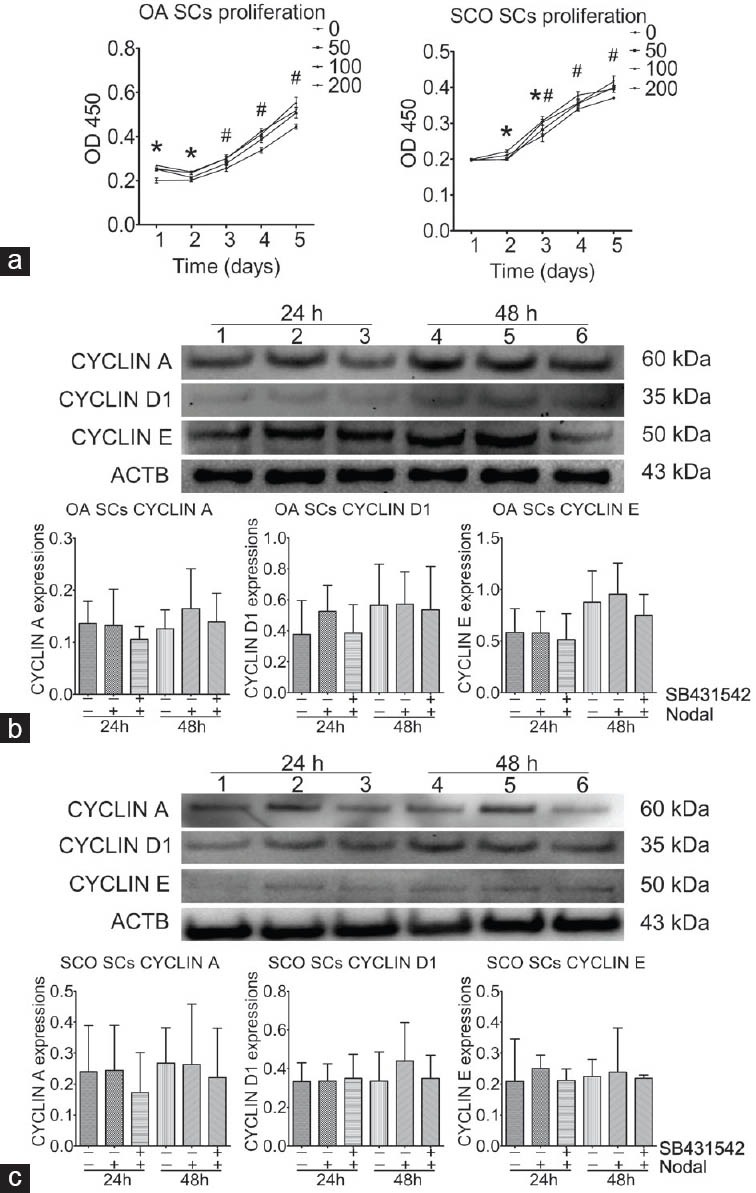
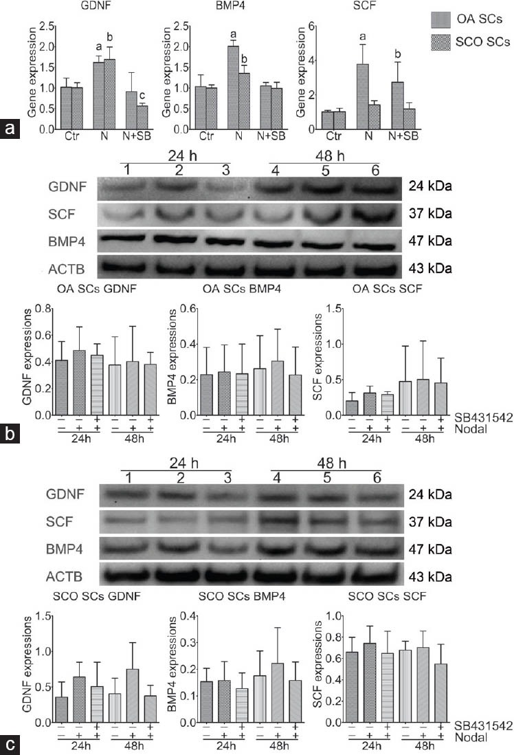
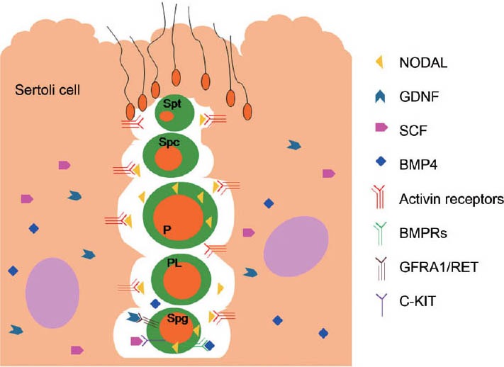
This study was designed to explore the regulatory effects of male germ cell secreting factor NODAL on Sertoli cell fate decisions from obstructive azoospermia (OA) and nonobstructive azoospermia (NOA) patients. Human Sertoli cells and male germ cells were isolated using two-step enzymatic digestion and SATPUT from testes of azoospermia patients. Expression of NODAL and its multiple receptors in human Sertoli cells and male germ cells were characterized by reverse transcription-polymerase chain reaction (RT-PCR) and immunochemistry. Human recombinant NODAL and its receptor inhibitor SB431542 were employed to probe their effect on the proliferation of Sertoli cells using the CCK-8 assay. Quantitative PCR and Western blots were utilized to assess the expression of Sertoli cell functional genes and proteins. NODAL was found to be expressed in male germ cells but not in Sertoli cells, whereas its receptors ALK4, ALK7, and ACTR-IIB were detected in Sertoli cells and germ cells, suggesting that NODAL plays a regulatory role in Sertoli cells and germ cells via a paracrine and autocrine pathway, respectively. Human recombinant NODAL could promote the proliferation of human Sertoli cells. The expression of cell cycle regulators, including CYCLIN A, CYCLIN D1 and CYCLIN E, was not remarkably affected by NODAL signaling. NODAL enhanced the expression of essential growth factors, including GDNF, SCF, and BMP4, whereas SB431542 decreased their levels. There was not homogeneity of genes changes by NODAL treatment in Sertoli cells from OA and Sertoli cell-only syndrome (SCO) patients. Collectively, this study demonstrates that NODAL produced by human male germ cells regulates proliferation and numerous gene expression of Sertoli cells.


Figures

Figure 1
Figure 1
Morphology of testis from OA and SCO patients and identification of the isolated human Sertoli cells, spermatogonia, spermatocytes, and spermatids. (a): H&E staining illustrated the morphology of testicular tissues from OA (left panel) and SCO patients (right panel). Histological examination showed that seminiferous tubule from SCO testis had a reduced diameter, with only Sertoli cells along the basement membrane, compared with that of OA. Scale bars = 10 μm. (b): The human Sertoli cells isolated from OA and SCO patient testes expressed transcripts of GATA4, ABP, WT1, and FSHR but no transcripts of VASA, HSD3B, CYP17A1, SMA, MYH11, CD34, and CD105. 1 and 2 represents OA Sertoli cells and SCO Sertoli cells, respectively. (c): RT-PCR showed that the expresion of specific makers in isolated human spermatogonia (GFRA1, RET, and UCHL1), spermatocytes (SCP1 and SCP3), and spermatids (PRM1, PRM2, TP1, and TP2). (d): Immunocytochemistry revealed that both GATA4 (red fluorescence) and WT1 (green fluorescence) were expressed in human Sertoli cells isolated from OA and SCO patients testes. The enlarged images were shown in the up-left of the pictures. Scale bar = 10 μm. (e): Immunocytochemistry displayed the co-expressions of GATA4 (red fluorescence) and VIMENTIN (green fluorescence) in human Sertoli cells isolated from OA and SCO patient testes. Scale bars = 10 μm. (f): Negtive control staining of immunocytochemistry of human Sertoli cells without primary antibody. Scale bars = 10 μm. (g): Immunocytochemistry displayed the co-expressions of GFRA1 (green fluorescence), PIWIL2 (green fluorescence), and ACROSIN (green fluorescence) with VASA (Red fluorescence) in isolated germ cells. (h): Negtive control staining of immunocytochemistry of the human germ cells without primary antibody. Scale bars = 10 μm. Notes – OA: obstructive azoospermia, SCO: Sertoli cell-only syndrome, SC: Sertoli cells; Spg: spermatogonia; Spc: spermatocytes; Spt: spermatids. Passage 2 Sertoli cells were used in this experiment.
Figure 2
Figure 2
Meiotic spread assays revealed the expression of SCP3 (red fluorescence), CREST (blue fluorescence), and MLH1 (green fluorescence) in the isolated spermatocytes. Scale bar = 10 μm.
Figure 3
Figure 3
Expression profiles of NODAL protein in OA and SCO testis. (a): Immunocytochemical staining showed that NODAL was expressed in VASA-positive cells in OA testis. No specific staining was seen in Sertoli cells and interstitium. There was no specific VASA and NODAL staining in SCO testis. There was nonspecific staining of the enclosed tubules and interstitium in both OA and SCO testis, which could also be seen in negative control slides (b and c). “*” indicated the basement membrane, while “^” indicated the spermatogonia. (b and c): Negative control of immunocytochemistry of the human testes. Notes – OA: obstructive azoospermia, SCO: Sertoli cell-only syndrome. Scale bars = 50 μm.
Figure 4
Figure 4
Expression profiles of NODAL's receptors in OA and SCO testis. (a): Immunohistochemical staining showed that ALK4, ALK7, and ACTR-IIB were detected in GATA4-positive Sertoli cells in OA testis. (a’): Negative control of immunocytochemistry of the OA testes, displaying nonspecific staining. (b): Immunohistochemical staining displayed that ALK4, ALK7, and ACTR-IIB were expressed in GATA4-positive Sertoli cells in SCO testis. No specific staining was seen in other cells. (b’): Negative control of immunohistochemistry of the OA testes without primary antibody. Scale bars = 50 μm. OA: obstructive azoospermia; SCO: Sertoli cell-only syndrome.
Figure 5
Figure 5
Expressions of NODAL and its multiple receptors in human Sertoli cells. (a): RT-PCR showed that the expressions of transcripts of NODAL and its receptors ALK4, ALK5, ALK7, ACTR-IIB, TDGF1, and CRYPTIC, in the isolated human Sertoli cells from OA and SCO patient testes. Passage 2 Sertoli cells were used in this experiment. (b and c): Immunocytochemistry revealed that ALK4 (red fluorescence), ALK7 (green fluorescence), and ACTR-IIB (green fluorescence) were detected in human Sertoli cells isolated from OA patient testes. No specific staining (c) was observed when cells were incubated with normal IgG instead of primary antibodies. (d and e): Immunocytochemistry revealed that ALK4 (red fluorescence), ALK7 (green fluorescence), and ACTR-IIB (green fluorescence) were expressed in human Sertoli cells isolated from SCO patient testes. No specific staining (e) was seen when cells were incubated with normal IgG instead of primary antibodies. The enlarged images were showed in the up-right of the merged pictures. Scale bars = 20 μm. OA: obstructive azoospermia; SCO: Sertoli cell-only syndrome; 1: OA Sertoli cells; 2: SCO Sertoli cells.
Figure 6
Figure 6
Expressions of NODAL's multiple receptors in isolated human spermatogonia, spermatocytes, and spermatids. (a): RT-PCR showed that the expression of NODAL's multiple receptors ALK4, ALK5, ALK7, ACTR-IIB, TDGF1, and CRYPTIC, in the isolated human spermatogonia, spermatocytes, and spermatids from OA patient testes. (b): Immunocytochemistry revealed that ALK4 (red fluorescence), ALK7 (green fluorescence), and ACTR-IIB (green fluorescence) were expressed in human spermatogonia isolated from OA patient testes. (c): Immunocytochemistry revealed that ALK4 (red fluorescence), ALK7 (green fluorescence), and ACTR-IIB (green fluorescence) were detected in human spermatocytes isolated from OA patient testes. (d): Immunocytochemistry revealed that ALK4 (red fluorescence), ALK7 (green fluorescence), and ACTR-IIB (green fluorescence) were expressed in human spermatids isolated from OA patient testes. Scale bars = 20 μm. OA: obstructive azoospermia; SCO: Sertoli cell-only syndrome.
Figure 7
Figure 7
Expression profiles of NODAL proteins in human germ cells. (a): Double staining showed that VASA (red fluorescence) and NODAL (green fluorescence) were co-expressed in human germ cells. (b): Immunohistochemistry revealed that NODAL (green fluorescence) was detected in spermatogonia (GFRA1: red fluorescence) and spermatocytes (PIWIL2: red fluorescence) but not in spermatids (ACROSIN: red fluorescence). (c): Negtive control of immunocytochemistry of the human germ cells, which showed no specific staining. Scale bars = 20 μm. Spg: spermatogonia; Spc: spermatocyte; Spt: spermatid.
Figure 8
Figure 8
The influence of NODAL in human Sertoli cell proliferation and cell cycle proteins. (a): CCK-8 assay showed that NODAL promoted human Sertoli cell growth. OD values of Sertoli cells under 100 ng ml−1 human recombinant NODAL treatment were remarkably higher than those of other groups from 24 h to 72 h of culture. “*” indicates that 100 ng ml−1 human recombinant NODAL treatment displayed the highest OD values when compared with those in other groups at the same time points (P < 0.05). “#” indicates that OD values of each group at that time point was higher than that of the 24 h of culture. (b and c), Western blots reflected the effect of NODAL on the expression of CYCLIN A, CYCLIN D1, and CYCLIN E in human Sertoli cells from OA and SCO patients. Notes: In b and c, 1, 2, and 3 represented proteins changes of OA and SCO Sertoli cells with the control, NODAL treatment, and NODAL and SB431542 treatment, respectively, for 24 h; 4, 5, and 6 represented proteins changes of OA and SCO Sertoli cells with control, NODAL treatment, and NODAL and SB431542 treatment, respectively, for 48 h. OA: obstructive azoospermia; SCO: Sertoli cell-only syndrome.
Figure 9
Figure 9
The effect of NODAL on the expression of several growth factors at transcription and translation levels in human Sertoli cells. (a): Real-time PCR showed the expression of GDNF, SCF, and BMP4 transcripts in OA Sertoli cells without or with NODAL and SB431542 treatment. “a” represented statistical difference compared with control group in OA Sertoli cell gene regulation. “b” represented statistical difference compared to control group in SCO Sertoli cell gene regulation (P < 0.05). (b and c): Western blots displayed the protein expression of GDNF, SCF, and BMP4 in human Sertoli cells without or with NODAL and SB431542. Notes: In a, Ctr: control group; N: NODAL group; N + SB: NODAL and SB431542 group. In b and c, 1, 2, and 3 represented protein changes of OA and SCO Sertoli cells with control, NODAL, or NODAL and SB431542 treatment, respectively, for 24 h; 4, 5, and 6 represented protein changes of OA and SCO Sertoli cells with control, NODAL, and NODAL and SB431542 treatment, respectively, for 48 h. OA: obstructive azoospermia; SCO: Sertoli cell-only syndrome.
Figure 10
Figure 10
Schematic diagram illustrated the cross-talk or interaction among male germ cells and Sertoli cells via NODAL signaling in human seminiferous tubules. NODAL is found to be secreted by spermatogonia and spermatocytes, which can exert its biological effect on regulating the proliferation and secretion of growth factors (e.g., GDNF, SCF, and BMP4) of human Sertoli cells. Sertoli cells can also produce other growth factors which play key roles in regulating spermatogenesis. The functional and appropriate interaction among male germ cells and Sertoli cells maintains the normal spermatogenesis. Spg: spermatogonia; PL: preleptene; P: pachytene spermatocyte; Spc: spermatocyte; Spt: spermatid.

Similar articles

Cited by

References

    1. Agarwal A, Tvrda E, Sharma R. Relationship amongst teratozoospermia, seminal oxidative stress and male infertility. Reprod Biol Endocrinol. 2014;12:45. - PMC - PubMed
    1. Ma M, Yang S, Zhang Z, Li P, Gong Y, et al. Sertoli cells from non-obstructive azoospermia and obstructive azoospermia patients show distinct morphology, Raman spectrum and biochemical phenotype. Hum Reprod. 2013;28:1863–73. - PubMed
    1. Johnston DS, Olivas E, DiCandeloro P, Wright WW. Stage-specific changes in GDNF expression by rat Sertoli cells: a possible regulator of the replication and differentiation of stem spermatogonia. Biol Reprod. 2011;85:763–9. - PMC - PubMed
    1. Sato T, Aiyama Y, Ishii-Inagaki M, Hara K, Tsunekawa N, et al. Cyclical and patch-like GDNF distribution along the basal surface of Sertoli cells in mouse and hamster testes. PLoS One. 2011;6:e28367. - PMC - PubMed
    1. Kanatsu-Shinohara M, Onoyama I, Nakayama KI, Shinohara T. Skp1-Cullin-F-box (SCF)-type ubiquitin ligase FBXW7 negatively regulates spermatogonial stem cell self-renewal. Proc Natl Acad Sci U S A. 2014;111:8826–31. - PMC - PubMed

Publication types

MeSH terms

Supplementary concepts