. 2015 Aug 20:6:8013.
doi: 10.1038/ncomms9013.

Structural mechanism for signal transduction in RXR nuclear receptor heterodimers

Affiliations

Structural mechanism for signal transduction in RXR nuclear receptor heterodimers

Douglas J Kojetin et al. Nat Commun. .

Abstract

A subset of nuclear receptors (NRs) function as obligate heterodimers with retinoid X receptor (RXR), allowing integration of ligand-dependent signals across the dimer interface via an unknown structural mechanism. Using nuclear magnetic resonance (NMR) spectroscopy, x-ray crystallography and hydrogen/deuterium exchange (HDX) mass spectrometry, here we show an allosteric mechanism through which RXR co-operates with a permissive dimer partner, peroxisome proliferator-activated receptor (PPAR)-γ, while rendered generally unresponsive by a non-permissive dimer partner, thyroid hormone (TR) receptor. Amino acid residues that mediate this allosteric mechanism comprise an evolutionarily conserved network discovered by statistical coupling analysis (SCA). This SCA network acts as a signalling rheostat to integrate signals between dimer partners, ligands and coregulator-binding sites, thereby affecting signal transmission in RXR heterodimers. These findings define rules guiding how NRs integrate two ligand-dependent signalling pathways into RXR heterodimer-specific responses.


Conflict of interest statement

Kendall Nettles is a consultant for Genentech.

Figures

Figure 1
Figure 1. NMR reveals a role for conformational dynamics in RXR permissiveness.
(a) CV-1 cells co-transfected with PPARγ expression plasmid, RXRα expression plasmid and a PPAR-responsive luciferase reporter. Cells were treated with vehicle, 10 μM 9cRA and/or 10 μM rosiglitazone for 24 h. Magenta and black bars are coloured to match NMR data in d, where ligand content is the same. Luciferase activity is shown normalized to vehicle-treated cells and was performed in quadruplicate, plotted with the average (±s.e.m) and representative of at least three experiments. (b) CV-1 cells co-transfected with TRβ expression plasmid, RXRα expression plasmid and a TR-responsive luciferase reporter. Cells were treated with vehicle, 100 nM LG100268 (LG268) and/or 1 μM T3 for 24 h. Luciferase activity is shown normalized to vehicle-treated cells and was performed in quadruplicate, plotted with the average (±s.e.m) and representative of at least two experiments. Magenta and black bars are coloured to match NMR data in e, where ligand content is the same. (c) Structural location of RXR residues mentioned in the NMR analysis. (d) Overlay of 2D [1H,15N]-TROSY-HSQC NMR data for [2H,13C,15N]-RXRα LBD in the apo form and coloured magenta—with the same bound to 9cRA and coloured black. (e) Overlay of 2D [1H,15N]-TROSY-HSQC NMR data for [2H,13C,15N]-RXRα LBD bound to 9cRA and heterodimerized to apo-PPARγ and coloured magenta—with the same bound to rosiglitazone (Rosi) and coloured black. (f) Overlay of 2D [1H,15N]-TROSY-HSQC NMR data for [2H,13C,15N]-RXRα LBD bound to 9cRA and heterodimerized to apo-TRβ and coloured magenta—with the same bound to T3 and coloured black.
Figure 2
Figure 2. Crystal structure of the TRβ·T3·SRC-2/apo-RXRα LBD complex.
(a) Structure of the TR/RXR LBD heterodimer is shown as ribbon with T3 as space filled. TR is coloured green, and the bound SRC-2 peptide is coloured blue and only binds to T3·TR. RXR is coloured light blue, with the dimer interface coloured coral and helix 12 coloured purple. There is no bound ligand in RXR, and RXR helix 12 adopts an inactive conformation positioned into the AF-2 co-activator-binding surface. (b) TR is superimposed on the RXR LBD homodimer (PDB 1MVC), showing conservation of domain structure. Structures are coloured as in a. (c) Same as b, except dimers are superimposed via the RXR protomer, rather than TR to RXR, illustrating the shift in the TR dimer interface relative to the other RXR promoter in the RXR homodimer. The magenta circle highlights the only region that superimposes similarly between TR and RXR, the amino-terminal end of TRβ helix 11.
Figure 3
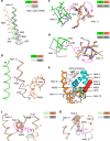
Figure 3. TR alters apo-RXR conformation via the helix 11 portion of the dimer interface.
(a) Helix 11 of the dimer interface shown as Cα traces for TRβ·T3·SRC-2/apo-RXRα, coloured green and coral, respectively. The RXR homodimer (PDB 1MVC), RXR/PPARγ (PDB 1FM6) and RXR/LXR (PDB 1UHL) heterodimers were superimposed on the TRβ·T3·SRC-2/apo-RXRα structure using the RXR promoter molecule and are coloured grey. (bd) The active conformation RXR homodimer (PDB 1MVC) superimposed on the TRβ·T3·SRC-2/apo-RXRα structure via RXR and coloured as in a, with the RXR homodimer coloured grey. (b) TR helix 11 (green) induces a shift in the RXR helix 11 (coral) relative to the RXR homodimer (grey). This shift is adjacent to RXR helix 5. (c) The unique position of TR T426 in helix 11 induces a shift in RXR P423 in helix 11 and a rotation of the RXR helix 11 backbone. (d) The location of TR A433 in helix 11 away from the dimer interface compared with the equivalent residue in RXR, L430, allows RXR L430 and the RXR helical backbone to rotate in context of the TRβ·T3·SRC-2/apo-RXRα heterodimer. (e) RXR in the active conformation (PDB 1MVC) with RXR ligand (MBS649) shown as space filled and SRC-2 peptide bound to the AF-2 co-activator-binding surface coloured red. RXR W305 in helix 5 mediates contacts with the ligand, M454 in helix 12 and the co-activator-binding site via L276 in helix 3. Colour is used to help differentiate secondary structural elements and provide depth for overlapping elements; helix 3 and 4 in cyan, helix 5 in blue, and helix 10/11 and helix 12 in orange. (f) TRβ·T3·SRC-2/apo-RXRα was superimposed with the active conformation RXR homodimer (PDB 1MVC) and shown as Cα trace. The rotation of helix 5 in TRβ·T3·SRC-2/apo-RXRα repositions W305 such that it clashes with the active conformation of RXR L276. (g) Same as f, but showing the active conformation of helix 12, and the clash with the rotated position of W305 in the TRβ·T3·SRC-2/apo-RXRα heterodimer.
Figure 4
Figure 4. Co-evolved amino acid network mediates structural allostery between RXR and TR.
(a) The TRβ·T3·SRC-2/apo-RXRα structure (green and coral) superimposed with the active conformation RXR homodimer (PDB 1MVC) via RXR and shown in grey. RXR residues in helix 11 (L425, R426) and helix 5 (W305, E307) are part of a network of co-evolved amino acids identified using a statistical coupling analysis (SCA). (b) The SCA network amino acids shown as space filled on the TRβ·T3·SRC-2/apo-RXRα heterodimer link the dimer interface, ligand-binding pocket and AF-2 co-activator-binding site. (c) Active conformation RXR homodimer (PDB 1MVC) superimposed with RXR from the TRβ·T3·SRC-2/apo-RXRα heterodimer. Shown are helix 3 and helix 4 of the AF-2 surface, and W305 in helix 5. The rotation of helix 5 induces an altered conformation of the AF-2 surface via the SCA network amino acids. (d) Regions in RXR that are protected from HDX on heterodimerizaton with TR.
Figure 5
Figure 5. Mutagenesis confirms a structure-function role for co-evolved amino acids.
(a,b) CV-1 cells co-transfected with TRβ expression plasmid, (a) RXRα or (b) RXRα L276V mutant expression plasmid, and a TR-responsive luciferase reporter. Cells were treated with vehicle, or the indicated dose of T3±1 μM 9cRA for 24 h. (c) Drosophila SL2 cells co-transfected with TRβ expression plasmid, RXRα or RXRα E434-mutant expression plasmid, a TR-responsive luciferase reporter. Cells were treated with vehicle, the RXR agonist LG100268 (LG268; 100 nM) and/or 1 μM TR agonist (T3) for 24 h. (d) CV-1 cells co-transfected with TRβ expression plasmid, RXRα or RXRα E434N mutant expression plasmid, and a TR-responsive luciferase reporter. Cells were treated with vehicle or the indicated dose of T3 overnight. (e) Drosophila SL2 cells co-transfected with VDR expression plasmid, RXRα or RXRα E434-mutant expression plasmid, and a VDR-responsive luciferase reporter. Cells were treated with vehicle, the RXR agonist LG100268 (LG268; 100 nM) and/or 1 μM VDR agonist (vitamin D3) for 24 h. Luciferase activity is shown normalized to vehicle-treated cells and was performed in quadruplicate; plotted with the average±s.e.m and representative of at least three experiments.
Figure 6
Figure 6. NMR reveals ligand binding to PPARγ affects the conformation of RXR.
NMR data are coloured grey for 9cRA-bound RXRα; black for 9cRA-bound RXRα heterodimerized to apo-PPARγ or the same bound to the following PPARγ ligands: rosiglitazone (magenta), MRL20 (blue), MRL24 (orange) or SR1664 (green); plotted on PPARγ/RXRα (PDB 1FM9). (a) NMR data (left) focusing on residues in RXRα helix 7 and helix 10/11 dimer interface that are perturbed by ligand binding to PPARγ, which are plotted onto the PPARγ/RXRα crystal structure and coloured according to structural location (yellow for helix 7; blue for helix 10/11); coloured dark if shown in the NMR data to the left or light if not. (b) NMR data (left) focusing on residues in core of RXRα that are perturbed by ligand binding to PPARγ, which are plotted onto the PPARγ/RXRα crystal structure and coloured red; and coloured dark if shown in the NMR data to the left or light if not. (c) NMR data (left) focusing on residues in RXRα helix 12, the AF-2 surface and the ligand-binding pocket that are perturbed by ligand binding to PPARγ, which are plotted onto the PPARγ/RXRα crystal structure and coloured according to structural location (green for AF-2/helix 12; orange for the ligand-binding pocket); coloured dark if shown in the NMR data to the left or light if not.
Figure 7
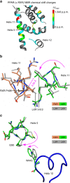
Figure 7. Co-evolved amino acid network with other receptors.
(a) NMR chemical shift perturbations in PPARγ on heterodimerization with RXRα mapped onto the structure of the PPARγ LBD (PDB 2PRG). (b) LXR homodimer (PDB 3IPU) coloured grey and superimposed with the LXR promoter from the LXR/RXR heterodimer (PDB 1UHL) coloured green and coral, respectively, shows that RXR induces rotation of LXR helix 11. (c) The RXR-induced shift in LXR helix 11 (PDB 1UHL) induces a rotation of LXR helix 5 relative to the LXR homodimer (PDB 3IPU), allowing W443 in helix 12 to adopt an alternative conformation with greater van der Waals contacts and increased buried surface area.
Figure 8
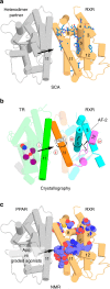
Figure 8. Summary of structural studies and allostery in RXR heterodimers.
(a) The SCA network residues plotted on RXR using the PPARγ/RXRα heterodimer structure as a model (PDB 1FM6). (b) Schematic diagram summarizing our TRβ·T3·SRC-2/apo-RXRα crystal structure showing how TR structurally silences RXR. The signal that emanates from TR (i) induces a shift in RXR helix 11 (ii), leading to a rotation of helix 5 (iii) resulting in structural arrangements that cause RXR helix 12 to adopt an inactive conformation (iv). (c) Summary of residues affected in the NMR analysis of ligand-selective signalling in PPARγ/RXRα, plotted on PDB 1FM6. Helix numbers are indicated for elements of interest. Arrows indicate the flow of the allosteric signal.

References

    1. Shulman A. I. & Mangelsdorf D. J. Retinoid x receptor heterodimers in the metabolic syndrome. N. Engl. J. Med. 353, 604–615 (2005). - PubMed
    1. Rosenfeld M. G., Lunyak V. V. & Glass C. K. Sensors and signals: a coactivator/corepressor/epigenetic code for integrating signal-dependent programs of transcriptional response. Genes Dev. 20, 1405–1428 (2006). - PubMed
    1. Shang Y. & Brown M. Molecular determinants for the tissue specificity of SERMs. Science 295, 2465–2468 (2002). - PubMed
    1. Choi J. H. et al. Anti-diabetic drugs inhibit obesity-linked phosphorylation of PPARgamma by Cdk5. Nature 466, 451–456 (2010). - PMC - PubMed
    1. Marciano D. P. et al. The therapeutic potential of nuclear receptor modulators for treatment of metabolic disorders: PPARgamma, RORs, and Rev-erbs. Cell Metab. 19, 193–208 (2014). - PubMed

Publication types

Associated data

LinkOut - more resources