. 2015 Aug 19;35(33):11543-58.
doi: 10.1523/JNEUROSCI.5267-14.2015.

Hepatocyte Growth Factor and MET Support Mouse Enteric Nervous System Development, the Peristaltic Response, and Intestinal Epithelial Proliferation in Response to Injury

Affiliations

Hepatocyte Growth Factor and MET Support Mouse Enteric Nervous System Development, the Peristaltic Response, and Intestinal Epithelial Proliferation in Response to Injury

Marina Avetisyan et al. J Neurosci. .

Abstract

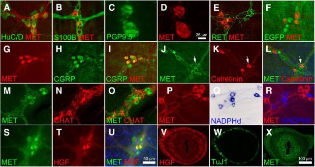
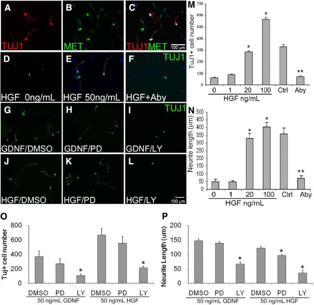
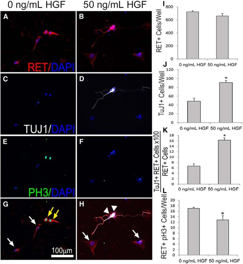
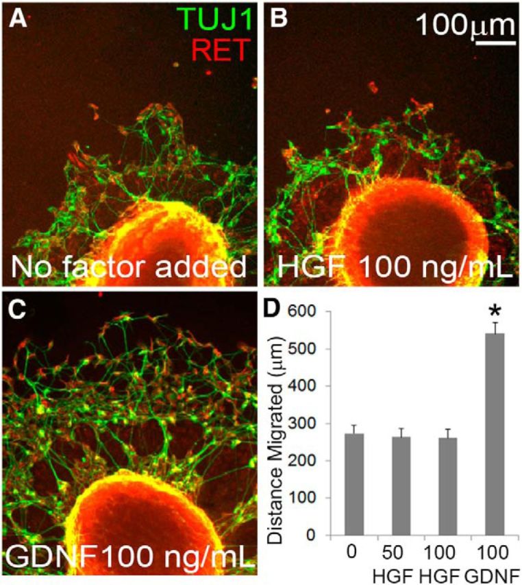
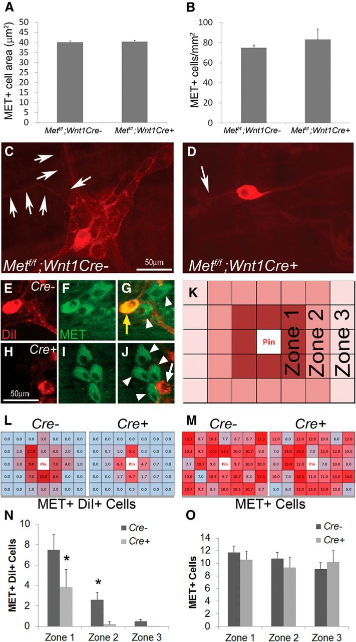
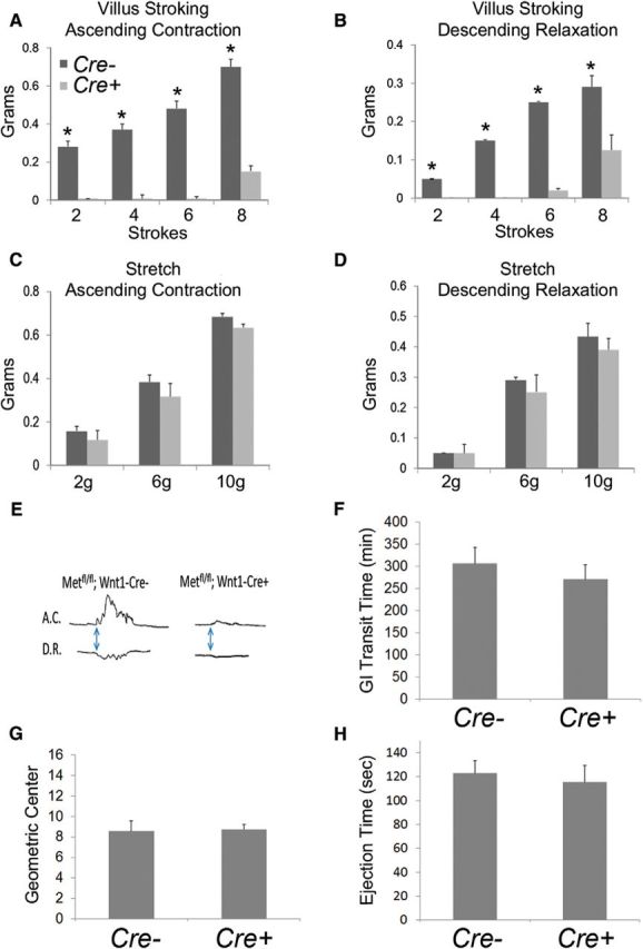
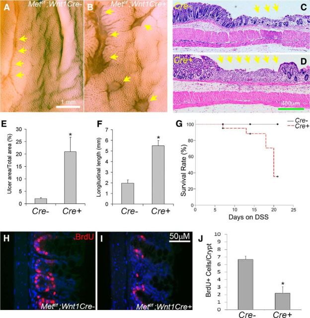
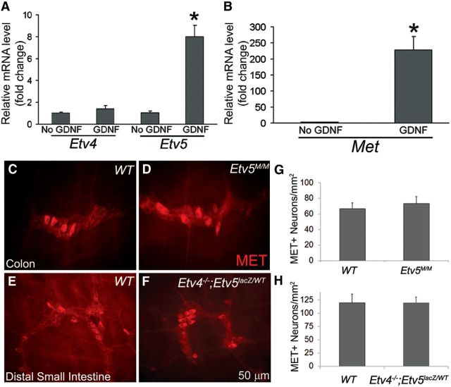
Factors providing trophic support to diverse enteric neuron subtypes remain poorly understood. We tested the hypothesis that hepatocyte growth factor (HGF) and the HGF receptor MET might support some types of enteric neurons. HGF and MET are expressed in fetal and adult enteric nervous system. In vitro, HGF increased enteric neuron differentiation and neurite length, but only if vanishingly small amounts (1 pg/ml) of glial cell line-derived neurotrophic factor were included in culture media. HGF effects were blocked by phosphatidylinositol-3 kinase inhibitor and by MET-blocking antibody. Both of these inhibitors and MEK inhibition reduced neurite length. In adult mice, MET was restricted to a subset of calcitonin gene-related peptide-immunoreactive (IR) myenteric plexus neurons thought to be intrinsic primary afferent neurons (IPANs). Conditional MET kinase domain inactivation (Met(fl/fl); Wnt1Cre+) caused a dramatic loss of myenteric plexus MET-IR neurites and 1-1'-dioctodecyl-3,3,3',3'-tetramethylindocarbocyamine perchlorate (DiI) labeling suggested reduced MET-IR neurite length. In vitro, Met(fl/fl); Wnt1Cre+ mouse bowel had markedly reduced peristalsis in response to mucosal deformation, but normal response to radial muscle stretch. However, whole-bowel transit, small-bowel transit, and colonic-bead expulsion were normal in Met(fl/fl); Wnt1Cre+ mice. Finally, Met(fl/fl); Wnt1Cre+ mice had more bowel injury and reduced epithelial cell proliferation compared with WT animals after dextran sodium sulfate treatment. These results suggest that HGF/MET signaling is important for development and function of a subset IPANs and that these cells regulate intestinal motility and epithelial cell proliferation in response to bowel injury.

Significance statement: The enteric nervous system has many neuronal subtypes that coordinate and control intestinal activity. Trophic factors that support these neuron types and enhance neurite growth after fetal development are not well understood. We show that a subset of adult calcitonin gene-related peptide (CGRP)-expressing myenteric neurons produce MET, the receptor for hepatocyte growth factor, and that loss of MET activity affects peristalsis in response to mucosal stroking, reduces MET-immunoreactive neurites, and increases susceptibility to dextran sodium sulfate-induced bowel injury. These observations may be relevant for understanding and treating intestinal motility disorders and also suggest that enhancing the activity of MET-expressing CGRP neurons might be a useful strategy to reduce bowel inflammation.

Keywords: MET; calcitonin gene-related peptide; dextran sodium sulfate (DSS); enteric nervous system; hepatocyte growth factor; intrinsic primary afferent neurons.


Figures

Figure 1.
Figure 1.
MET immunoreactivity was detected in a subset of myenteric plexus CGRP+ IPANs. A, MET immunoreactivity was detected in 34 ± 6% of HuC/D+ myenteric neurons of the adult mouse small bowel. All MET+ cells expressed the pan-neuronal marker HuC/D. B, MET staining was absent from S100B+ enteric glia. C, D, MET was detected in human myenteric neurons using colon cross sections. E, F, MET and RET are detected in mutually exclusive sets of neurons as confirmed by immunohistochemistry (E) and by using a RET-EGFP reporter mouse (F). G–I, 100% of MET+ neurons were CGRP+ and 50% of CGRP+ neurons were MET+. J–L, MET was also found in 16 ± 5% of calretinin+ cells and 8 ± 2% of MET+ cells are calretinin+. Arrows highlight a MET+ calretinin+ neuron. M–O, 12 ± 0.1% of ChAT+ cells were MET+ and 8 ± 0.1% of MET+ neurons were ChAT+. P–R, There was no overlap between MET and NADPH diaphorase-stained nitric oxide-producing neurons. S–U, HGF was detected in 43 ± 11% of MET+ neurons and 100% of the HGF+ neurons were MET+. V, In cross sections of E14.5 fetal bowel, HGF-IR was found in mesenchymal cells surrounding developing ENS stained with TuJ1 (W). X, At E14.5, MET+ cells were present in the region of developing ENS as well as in gut epithelium and mesenchymal cells. Scale bars in U applies to A, B, E–U. Scale bar in D applies to C and D. Scale bar in X applies to V–X. N ≥ 3 replicates/staining condition.
Figure 2.
Figure 2.
HGF promoted neurogenesis and neurite growth in cultured E12.5 ENS precursor cells. A–C, E12.5 ENS precursors immunoselected with p75NTR antibody were maintained in culture for 48 h in the presence of HGF plus 1 pg/ml GDNF before TuJ1 and/or MET immunohistochemistry and DAPI nuclear staining. All TuJ1+ enteric neurons were MET-IR. D–F, M, N, HGF caused a dose-dependent increase in TuJ1-IR neuron number and neurite length. *p < 0.05, ANOVA with Dunn's multiple-comparison test. F, M, N, MET blocking antibody (Aby) reduced TuJ1+ neuron number and neurite length in surviving cells. Control (Ctrl) is 50 ng/ml HGF plus 1 pg/ml GDNF. **p < 0.01, Student's t test. G–L, O, P, When ENS precursors were grown in GDNF alone, the MEK inhibitor PD98059 (PD) had no effect on neuron number or neurite length, but the PI-3K inhibitor LY294002 (LY) reduced neuron number and neurite length. In contrast, in HGF (50 ng/ml) plus GDNF (1 pg/ml)-treated cells, both MEK and PI-3K inhibition reduced neurite length (P), whereas only PI-3K inhibition reduced neuron number (O). *p < 0.01, ANOVA with Dunn's multiple-comparison test. Scale bar in C applies to A–F. Scale bar in L applies to G–L. (N ≥ 3 biological replicates/group; 12 individual wells/group).
Figure 3.
Figure 3.
HGF/MET signaling enhanced ENS precursor differentiation into neurons in vitro. A–H, E12.5 ENS precursors were maintained in culture after p75NTR immunoselection for 2 d in the presence or absence of 50 ng/ml HGF before immunohistochemistry using RET (A, B), TuJ1 (C, D), and pH3 (E, F) antibodies as well as DAPI nuclear staining (A–H). G, H, Merged images. I–K, While the total number of RET+ cells was not altered by HGF (I), the total number of TuJ1+ neurons (J) and the percentage of RET+ cells that were TuJ1+ (K) increased with HGF treatment. L, The number of dividing precursor cells (pH3 and RET double positive) decreased with HGF treatment, suggesting that HGF increased neuronal differentiation and decreased proliferation. White arrows, Nonmitotic RET+ pH3− ENS precursors. Yellow arrow, Mitotic RET+PH3+ ENS precursors. White arrowhead, RET+TuJ1+ neurons. Scale bar in G applies to all images (N = 3 biological replicates/group; 12 individual wells/group; *p < 0.01, Student's t test).
Figure 4.
Figure 4.
HGF/MET signaling did not increase ENS precursor migration in culture. A–C, E12.5 gut slices were cultured 24 h on fibronectin-coated dishes with no added factor, HGF, or GDNF before staining for RET and TuJ1. A, ENCDCs migrate from the gut slice onto the culture dish even without any added factors. B, HGF did not increase the distance that ENCDCs migrated from the edge of the gut slice. C, GDNF markedly increased the distance ENCDCs migrate from the edge of the gut slice. D, Quantitative data (no added factor, 15 slices; 50 ng/ml HGF, 14 slices; 100 ng/ml HGF, 19 slices; 100 ng/ml GDNF, 30 slices; N = 3 independent experiments). *p < 0.001 for GDNF versus no added factor, ANOVA with Dunn's multiple-comparison test.
Figure 5.
Figure 5.
MET deletion within ENS precursors caused selective defects in MET-expressing enteric neurons in adult mice. A, B, The myenteric plexus of mice lacking functional MET receptors in the ENS (Mettf/f; Wnt1Cre+) contained MET+ neuronal cell bodies of normal size and abundance (N = 3 mice of each genotype/condition). C, D, Mettf/f; Wnt1Cre+ mice had few interganglionic and intraganglionic MET-IR neurites (N = 6 mice of each genotype/condition). E–J, Double-labeling for myenteric neurons combining DiI labeling and MET immunohistochemistry. Cells shown were two 20× fields away from the DiI application site. Yellow arrow, DiI+ MET+ cell body. White arrow, DiI+ MET− cell body. White arrowheads, DiI− MET+ cell bodies (N = 4 mice of each genotype; N > 8 distal small-bowel DiI-labeled regions/genotype). K, Schematic for analysis of DiI/MET-labeled samples: tissue pieces were divided into a 5 × 7 grid of 20× fields centered on the pin insertion site. The grid was additionally subdivided into three zones of varying distances from the pin. L, M, Heat map representations of the average number of MET+ DiI+ cells (L) and total MET+ cells (M) in each 20× field of the grid. Dark red > light red > blue for the number of cells in each region. N, The number of DiI+ MET+ cell bodies was dramatically reduced in Met cKO mice compared with controls in Zones 1 and 2. *p < 0.02 (Student's t test). O, The number of total MET+ cell bodies did not differ at varying distances from the pin site.
Figure 6.
Figure 6.
MET deletion within ENS precursors resulted in selective defects in the sensory arm of the peristaltic response. A, B, E, Mice lacking functional MET receptors in the ENS (Metfl/fl; Wnt1 Cre+) had an abnormal peristaltic reflex in response to mechanical stimulation of the villi, as evidenced by a severely blunted ascending contraction (A) and descending relaxation (B). C, D, In contrast, the peristaltic reflex elicited by circular muscle stretch had normal ascending contraction (C) and descending relaxation (D) in Metfl/fl;Wnt1 Cre+ mice, suggesting a selective sensory defect (N = 3 mice/genotype; *p < 0.01, Student's t test). F, Whole gastrointestinal transit as measured by the time needed to pass orally gavage-fed carmine dye in stool was not altered in Metfl/fl;Wnt1 Cre+ mice (N = 8 Met cKO and 7 control mice). G, Small-bowel transit as measured by determining the geometric center of FITC-dextran within the bowel 90 min following oral gavage was not altered in Metfl/fl;Wnt1 Cre+ mice (N = 4 Met cKO and 4 control mice). H, Colonic transit, as measured by the time taken to expel a bead placed 2 cm into the distal colon was not altered in Metfl/fl;Wnt1 Cre+ mice (N = 8 Met cKO and 7 control mice).
Figure 7.
Figure 7.
MET inactivation in ENS precursors increased mucosal injury in response to DSS treatment. A–F, Metfl/fl; Wnt1Cre+ mice and Metfl/fl; Wnt1Cre control animals were treated with 2.5% DSS in drinking water for 14 d and then examined using a dissecting microscope (A, B) or after paraffin sectioning and hematoxylin and eosin staining (C, D). E, F, Quantitative analysis of ulcer area in the descending colon and ulcer length in the rectum demonstrated increased ulcers in Metf/f; Wnt1Cre+ mice compared with controls. *p < 0.01, Student's t test. G, Kaplan–Meier analysis demonstrated that DSS-treated Metfl/fl; Wnt1Cre+ mice had higher death rates than controls (N = 8 Met cKO and 11 control mice). p < 0.05, log-rank test. H–J, BrdU labeling after 7 d of DSS treatment showed reduced colonic epithelial cell proliferation within crypts of Metf/f; Wnt1Cre mice compared with control animals. *p < 0.01, Student's t test. Yellow arrows: ulcerated regions (N = 5 Met cKO and 4 control animals/group for ulcer analysis and BrdU labeling).
Figure 8.
Figure 8.
GDNF/RET signaling increased Met and Etv5 mRNA in cultured ENS precursors, but MET protein immunoreactivity appeared normal in mice with Etv4 and Etv5 mutations. A, ENS precursors grown with 1 pg/ml GDNF had an eightfold increase in Etv5 mRNA, but not in Etv4 mRNA when compared with precursors cultured without GDNF. B, ENS precursors grown with 1 pg/ml GDNF also had a 230-fold increase in Met mRNA (N = 3 biological replicates). C–H, The myenteric plexus of adult Etv5M/M mice and P14 Etv4−/−; Etv5lacZ/WT compound mutants appeared grossly normal with no differences in MET-IR neuron density (N = 3 mice for each group). *p < 0.01 (Student's t test).

Similar articles

Cited by

References

    1. Akimoto M, Baba A, Ikeda-Matsuo Y, Yamada MK, Itamura R, Nishiyama N, Ikegaya Y, Matsuki N. Hepatocyte growth factor as an enhancer of nmda currents and synaptic plasticity in the hippocampus. Neuroscience. 2004;128:155–162. doi: 10.1016/j.neuroscience.2004.06.031. - DOI - PubMed
    1. Arthur LG, Schwartz MZ, Kuenzler KA, Birbe R. Hepatocyte growth factor treatment ameliorates diarrhea and bowel inflammation in a rat model of inflammatory bowel disease. J Pediatr Surg. 2004;39:139–143. discussion 139–143. - PubMed
    1. Baliu-Piqué M, Jusek G, Holzmann B. Neuroimmunological communication via CGRP promotes the development of a regulatory phenotype in TLR4-stimulated macrophages. Eur J Immunol. 2014;44:3708–3716. doi: 10.1002/eji.201444553. - DOI - PubMed
    1. Bischoff SC, Mailer R, Pabst O, Weier G, Sedlik W, Li Z, Chen JJ, Murphy DL, Gershon MD. Role of serotonin in intestinal inflammation: knockout of serotonin reuptake transporter exacerbates 2,4,6-trinitrobenzene sulfonic acid colitis in mice. Am J Physiol Gastrointest Liver Physiol. 2009;296:G685–G695. doi: 10.1152/ajpgi.90685.2008. - DOI - PubMed
    1. Bjerknes M, Cheng H. Modulation of specific intestinal epithelial progenitors by enteric neurons. Proc Natl Acad Sci U S A. 2001;98:12497–12502. doi: 10.1073/pnas.211278098. - DOI - PMC - PubMed

Publication types