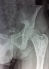
Myxoma of the femur: an unusual site of origin
- PMID: 26290566
- PMCID: PMC4550970
- DOI: 10.1136/bcr-2015-211480
Myxoma of the femur: an unusual site of origin
Abstract
Skeletal myxomas are rare benign tumours. Their occurrence in long bones of the extremities is rarely reported. A 45-year-old man presented with pain in his left proximal thigh for a duration of 4 months. Movements of the hip were painful. Radiography revealed an expansile osteolytic lesion in the left proximal femur near the lesser trochanteric region. On MRI, the lesion showed a homogenous signal enhancement with no cortical disruption. Extended curettage and bone grafting was performed. On gross examination, the curetted specimen was a yellowish-white mucoid material. Histopathology showed a tumour consisting of spindle-shaped and stellate-shaped cells with widely separated myxoid mucoidy stroma, suggestive of intraosseous myxoma. At 2 years follow-up, there were no signs of recurrence and the patient was doing well with excellent hip and knee function.
2015 BMJ Publishing Group Ltd.
Figures


References
-
- McClure DK, Dahlin DC. Myxoma of bone: report of three cases. Mayo Clin Proc 1977;52:249–53. - PubMed
-
- Hill JA, Victor TA, Dawson WJ et al. . Myxoma of toe. J Bone Joint Surg Am 1978;60:128–30. - PubMed
-
- Chaha PB, Tan KK. Periosteal myxoma of femur. J Bone Joint Surg Am 1972;54:1091–4. - PubMed
-
- Bauer WH, Harell A. Myxoma of bone. J Bone Joint Surg Am 1954;36(A:2):263–6. - PubMed
Publication types
MeSH terms
LinkOut - more resources
Full Text Sources
Other Literature Sources
Medical
