. 2015 Aug 4:3:e1115.
doi: 10.7717/peerj.1115. eCollection 2015.

Awake fMRI reveals a specialized region in dog temporal cortex for face processing

Affiliations

Awake fMRI reveals a specialized region in dog temporal cortex for face processing

Daniel D Dilks et al. PeerJ. .

Abstract

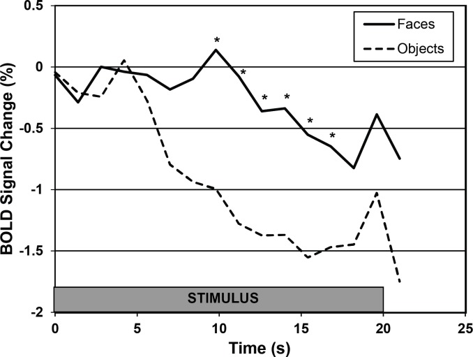
Recent behavioral evidence suggests that dogs, like humans and monkeys, are capable of visual face recognition. But do dogs also exhibit specialized cortical face regions similar to humans and monkeys? Using functional magnetic resonance imaging (fMRI) in six dogs trained to remain motionless during scanning without restraint or sedation, we found a region in the canine temporal lobe that responded significantly more to movies of human faces than to movies of everyday objects. Next, using a new stimulus set to investigate face selectivity in this predefined candidate dog face area, we found that this region responded similarly to images of human faces and dog faces, yet significantly more to both human and dog faces than to images of objects. Such face selectivity was not found in dog primary visual cortex. Taken together, these findings: (1) provide the first evidence for a face-selective region in the temporal cortex of dogs, which cannot be explained by simple low-level visual feature extraction; (2) reveal that neural machinery dedicated to face processing is not unique to primates; and (3) may help explain dogs' exquisite sensitivity to human social cues.

Keywords: Dog; Face area; fMRI.


Conflict of interest statement

Mark Spivak is president of Comprehensive Pet Therapy. Gregory Berns and Mark Spivak own equity in Dog Star Technologies and developed technology used in the research described in this paper. The terms of this arrangement have been reviewed and approved by Emory University in accordance with its conflict of interest policies.

Figures

Figure 1
Figure 1. Experimental setup in MRI.
Dogs were trained to station within an individually customized chin rest placed inside a stock human neck coil. The upper surface coil was located just superior to the dog’s head. Images were rear projected onto a translucent screen placed at the end of the magnet bore. In the dynamic stimuli runs, color movie clips (3-s each) were shown in 21 s blocks of human faces, objects (toys), scenes, and scrambled objects. In the static stimuli runs, black and white images (600 ms on, 400 ms off) were shown in 20 s blocks of human faces, dog faces, everyday objects, scenes, and scrambled faces. The dynamic stimuli runs were used to localize a candidate face region in the temporal cortex of dogs, and then the static stimuli runs were used to independently test the face selectivity of this region.
Figure 2
Figure 2. ROI locations for the dog face area (DFA) and primary visual cortex (V1).
The DFA was identified by the contrast of faces versus objects during the dynamic stimuli runs. Each color represents the ROI of one dog. For visualization and comparison of location, the ROIs have been spatially normalized and overlaid on a high resolution dog brain atlas (Datta et al., 2012). The location of the DFA was localized to the medial bank of the ventrocaudal temporal lobe in 4 of the 6 dogs, with the other 2 localized more laterally. V1 was identified by the average of all dynamic run conditions (face, objects, scenes, scrambled) relative to baseline. In each dog, a dorsal area of activation in the caudal portions of the marginal/endomarginal gyri was identified and corresponded to the known location of primary visual cortex.
Figure 3
Figure 3. Average percent signal change in DFA and V1.
Error bars indicate the standard error of the mean (n = 6). (A) In DFA, we found a significant category effect (F(3, 24) = 3.79, p = 0.02), with a significantly greater response to images of faces compared to objects (∗∗ p = 0.004) and a marginally greater response to scenes (p = 0.06). (B) V1 had a similar level of response to all stimulus categories (F(3, 24) = 0.42, p = 0.74), and crucially was significantly different from DFA in face selectivity (i.e., faces compared to objects) (F(1, 30) = 6.68, p = 0.02).
Figure 4
Figure 4. Average time course of activation in DFA for faces and objects.
The stimulus was visible for 20 s. Each time course was referenced to the volume immediately preceding the onset of the stimulus and was averaged over all dogs and all trials (excluding censored volumes). The response to objects decayed quickly while the response to faces was sustained, resulting in an overall greater response, which was individually significant at the indicated time points ( t > 1.65).

References

    1. Beitz AJ, Fletcher TF. The brain. In: Evans HE, editor. Miller’s anatomy of the dog. Third Edition. Philadelphia: W.B. Saunders Company; 1993.
    1. Berns GS, Brooks AM, Spivak M. Functional MRI in awake unrestrained dogs. PLoS ONE. 2012;7:e1115. doi: 10.1371/journal.pone.0038027. - DOI - PMC - PubMed
    1. Berns GS, Brooks A, Spivak M. Replicability and heterogeneity of awake unrestrained canine fMRI responses. PLoS ONE. 2013;8:e1115. doi: 10.1371/journal.pone.0081698. - DOI - PMC - PubMed
    1. Bruce V, Young A. In the eye of the beholder: the science of face perception. New York: Oxford University Press; 1998.
    1. Cook PF, Spivak M, Berns GS. One pair of hands is not like another: caudate BOLD response in dogs depends on signal source and canine temperament. PeerJ. 2014;2:e1115. doi: 10.7717/peerj.596. - DOI - PMC - PubMed