

Review
doi: 10.1016/j.neuron.2015.07.033.
Excitatory/Inhibitory Balance and Circuit Homeostasis in Autism Spectrum Disorders
Affiliations
- PMID: 26291155
- PMCID: PMC4567857
- DOI: 10.1016/j.neuron.2015.07.033
Item in Clipboard
Review
Excitatory/Inhibitory Balance and Circuit Homeostasis in Autism Spectrum Disorders
Neuron.
.
Abstract
Autism spectrum disorders (ASDs) and related neurological disorders are associated with mutations in many genes affecting the ratio between neuronal excitation and inhibition. However, understanding the impact of these mutations on network activity is complicated by the plasticity of these networks, making it difficult in many cases to separate initial deficits from homeostatic compensation. Here we explore the contrasting evidence for primary defects in inhibition or excitation in ASDs and attempt to integrate the findings in terms of the brain's ability to maintain functional homeostasis.
Copyright © 2015 Elsevier Inc. All rights reserved.
Figures
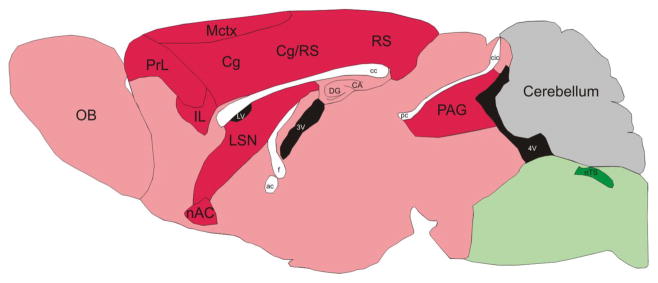
Low Fos expression in the motor cortex (Mctx) and adjacent regions indicates decreased activity while there is higher Fos expression in the nucleus of the solitary tract (nTS) and nearby areas. Differences in Fos expression in the Mecp2 null brain compared to wild-type are color coded as follows: Red, Null < Wt; Green, Null > Wt. Reproduced with permission from (Kron et al., 2012).
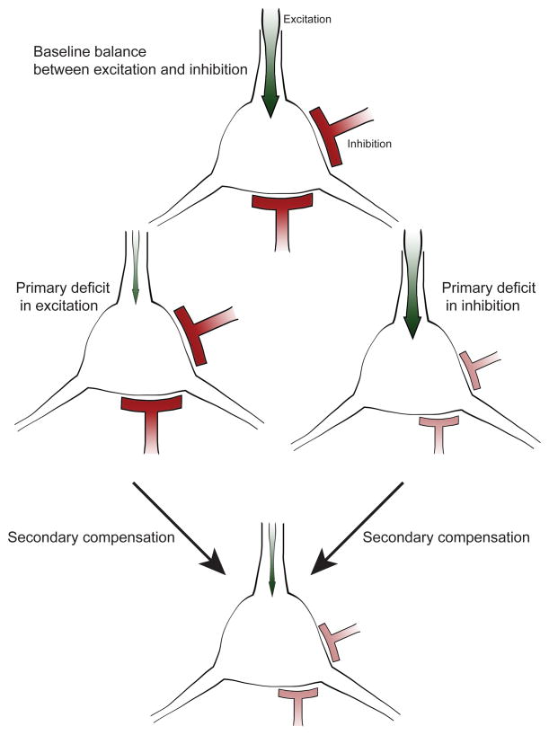
Proper neural network function relies on the balance between excitatory (green) and inhibitory (red) input. Primary defects in excitation or inhibition can be corrected via secondary compensatory mechanisms to restore balance and maintain network function. When a cell receives reduced excitation, secondary mechanisms down regulate the amount of inhibitory input onto this cell. Similarly, the excitatory input is decreased in response to a deficit in inhibition. Hence changes in both classes of synapses can appear similar following disease mechanisms that initially affect only one or the other.
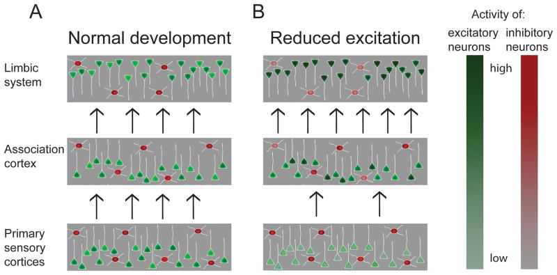
Cortical networks can be schematized as interconnected layers of neurons. Activity in the “input layer” of primary sensory cortices is driven by sensory inputs, while activity in higher order association and limbic regions depends to a greater degree on activity in preceding layers. (A) During normal development excitation and inhibition are balanced to preserve appropriate activity levels across synaptically connected brain regions with the activity of the cells in each layer adjusted to the amount of input this layer receives. (B) If the balance is perturbed so that, for example, input layers have reduced activity (indicated by normal red inhibitory but reduced green excitatory activity), homeostatic mechanisms compensate for the defect and upregulate the excitability of circuits in higher order Association and Limbic regions (indicated by a darker shade of green and lighter shade of red in some neurons) in an attempt to maintain normal levels of propagating activity. However, if not perfectly balanced, this can lead to overactivity in higher order regions coexisting with reduced activity in lower order regions. Networks in higher order regions with enhanced excitation and reduced inhibition may be brittle and prone to develop epileptiform activity.
References
-
- Asaka Y, Jugloff DG, Zhang L, Eubanks JH, Fitzsimonds RM. Hippocampal synaptic plasticity is impaired in the Mecp2-null mouse model of Rett syndrome. Neurobiol Dis. 2006;21:217–227. - PubMed
-
- Barros CS, Calabrese B, Chamero P, Roberts AJ, Korzus E, Lloyd K, Stowers L, Mayford M, Halpain S, Müller U. Impaired maturation of dendritic spines without disorganization of cortical cell layers in mice lacking NRG1/ErbB signaling in the central nervous system. PNAS. 2009;106:4507–4512. - PMC - PubMed
Publication types
MeSH terms
Grants and funding
LinkOut - more resources
Full Text Sources
Other Literature Sources
