. 2015 Dec;18(12):1317-26.
doi: 10.1089/jmf.2015.3443. Epub 2015 Aug 20.

Ilex latifolia Prevents Amyloid β Protein (25-35)-Induced Memory Impairment by Inhibiting Apoptosis and Tau Phosphorylation in Mice

Affiliations

Ilex latifolia Prevents Amyloid β Protein (25-35)-Induced Memory Impairment by Inhibiting Apoptosis and Tau Phosphorylation in Mice

Joo Youn Kim et al. J Med Food. 2015 Dec.

Abstract

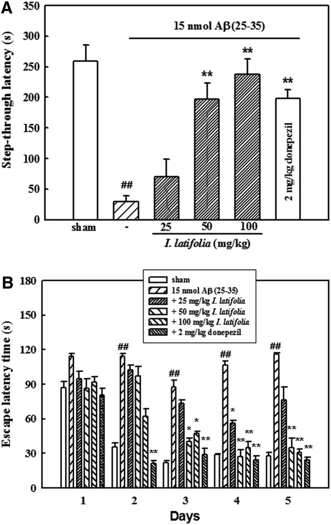
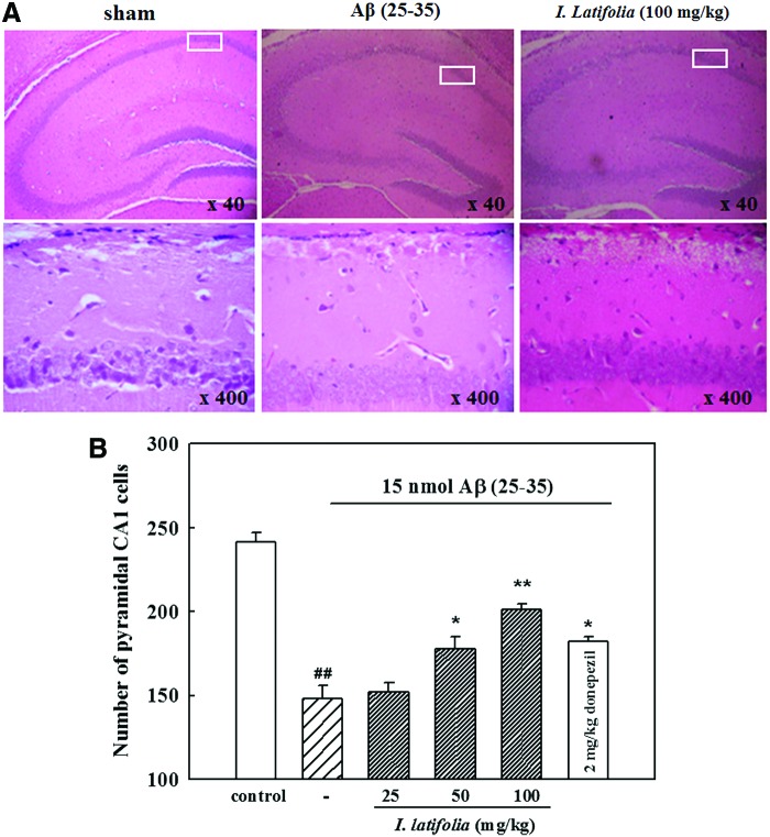
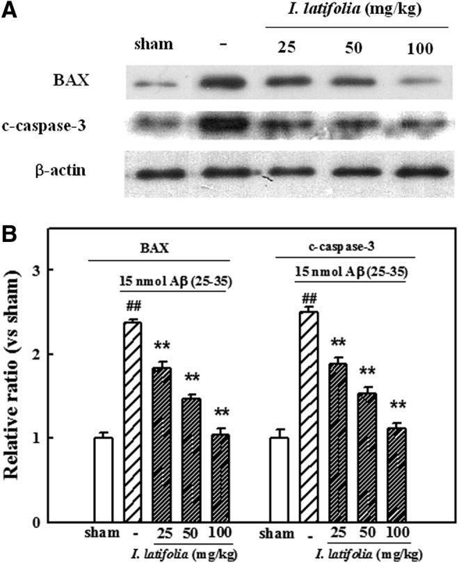
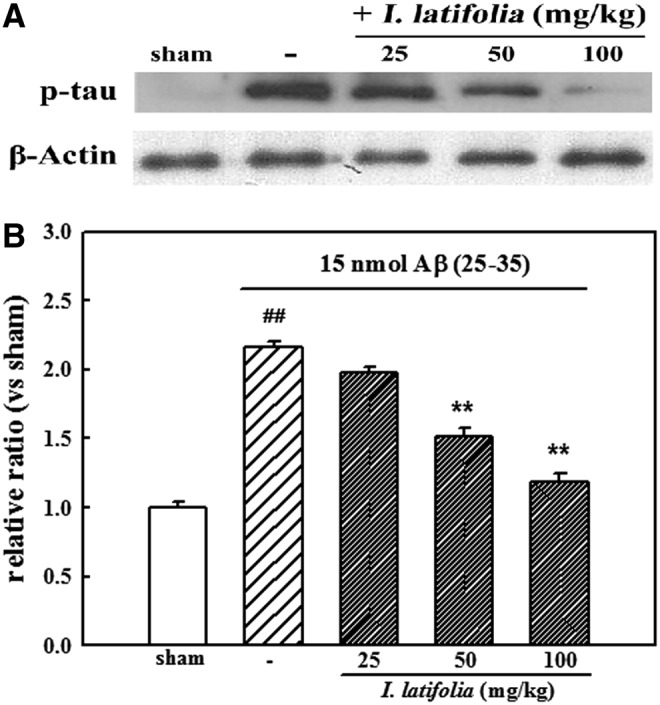
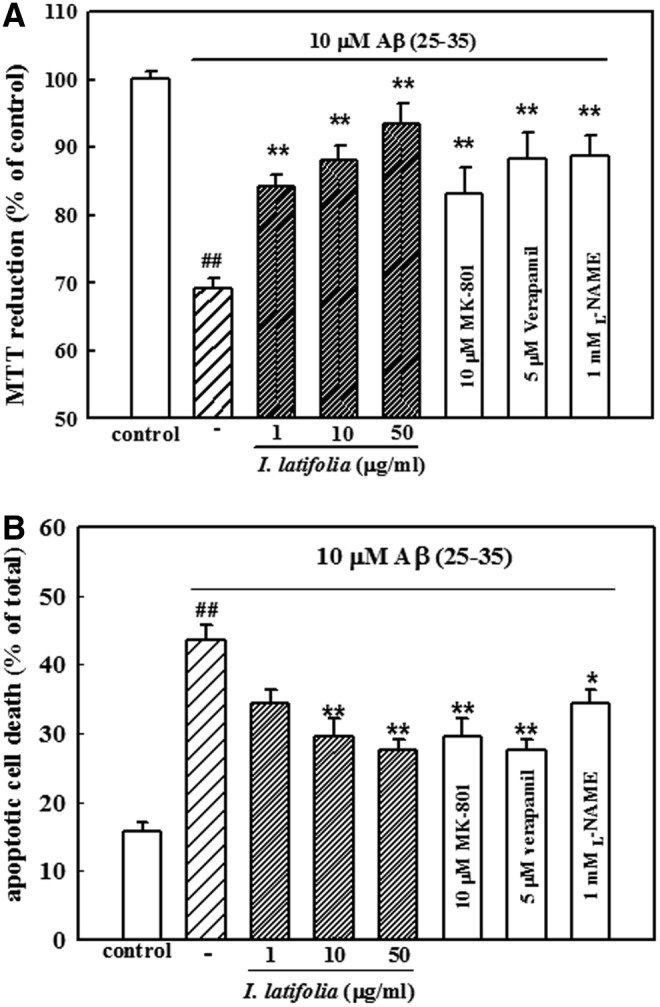
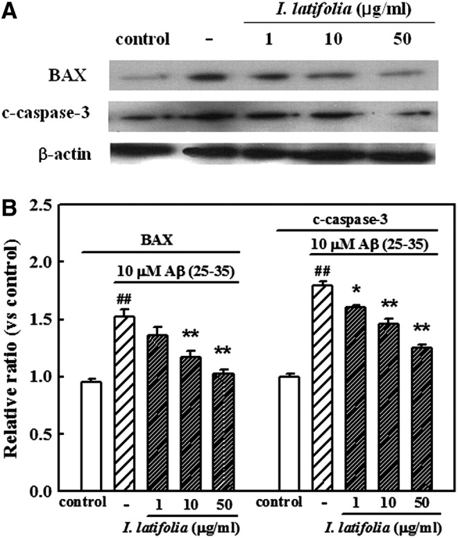
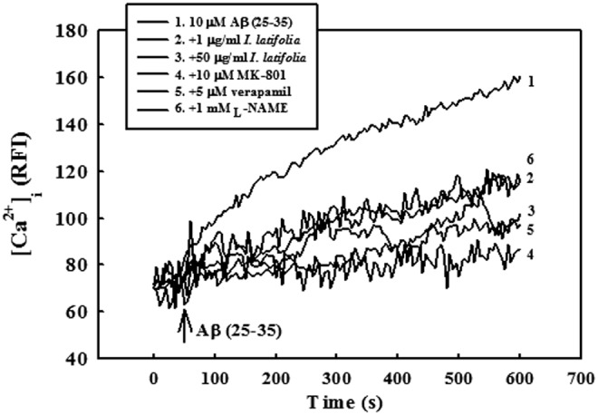
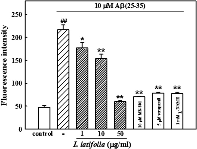
Ilex latifolia Thunb. (Aquifoliaceae), a Chinese bitter tea called "kudingcha," has been widely consumed as a health beverage and found to possess antioxidant, antidiabetic, antihypertensive, anti-inflammatory, and anti-ischemic activities. The aim of the present study was to investigate the neuroprotective effects of an ethanol extract of I. latifolia against amyloid β protein (Aβ)-induced memory impairment in mice and neurotoxicity in cultured rat cortical neurons. Memory impairment in mice was induced by intracerebroventricular injection of 15 nmol Aβ (25-35) and measured by the passive avoidance test and Morris water maze test. Chronic administration of I. latifolia (25-100 mg/kg, p.o.) significantly prevented Aβ (25-35)-induced memory loss. I. latifolia also prevented the decrease of glutathione concentrations, increased lipid peroxidation, expression of phosphorylated tau (p-tau), and changes in apoptosis-associated proteins in the memory-impaired mouse brain. Exposure of cultured cortical neurons to 10 μM Aβ (25-35) for 36 h induced neuronal apoptotic death. The neuronal cell death, elevation of intracellular Ca(2+) concentration, generation of reactive oxygen species, and expression of proapoptotic proteins caused by Aβ (25-35) in the cultured neurons were inhibited by treatment with I. latifolia (1-50 μg/mL). These results suggest that I. latifolia may have a possible therapeutic role in managing cognitive impairment associated with Alzheimer's disease. The underlying mechanism might involve the antiapoptotic effects mediated by antioxidant activity and inhibition of p-tau formation.

Keywords: Alzheimer's disease; Ca2+ signaling; antioxidant; cognition; neuroprotection.


Figures

<b>FIG. 1.</b>
FIG. 1.
Protective effects of I. latifolia against amyloid β protein (Aβ) (25–35)-induced memory impairment in mice. Learning and memory performance of the mice were assessed by (A) a passive avoidance test (n = 11–13) and (B) a Morris water maze test (n = 8–10). Values are expressed as the mean ± standard error of the mean (SEM) ##P < .01 versus the sham; *P < .05 and **P < .01 versus 15 nmol Aβ (25–35).
<b>FIG. 2.</b>
FIG. 2.
Representative photomicrographs of (A) the hippocampus (upper) and CA1 region (lower), and (B) histogram of CA1 pyramidal cell numbers illustrating the inhibitory effect of I. latifolia on Aβ (25–35)-induced hippocampal damage in mice. Brain sections were stained with hematoxylin and eosin. Pathological features of the brain sections 7 days after intracerebroventricular injection of 15 nmol Aβ (25–35) were observed using a brightfield microscope. ##P < .01 versus sham; *P < .05 and **P < .01 versus 15 nmol Aβ (25–35). Color images available online at www.liebertpub.com/jmf
<b>FIG. 3.</b>
FIG. 3.
Inhibitory effect of I. latifolia on Aβ (25–35)-induced expression of Bax and c-caspase-3 in mice brains. (A) Representative Western blot of proteins in the brains. (B) Bar graphs showing the relative ratio of Bax and c-caspase-3 proteins compared to β-actin. ##P < .01 versus the sham; **P < .01 versus 15 nmol Aβ (25–35).
<b>FIG. 4.</b>
FIG. 4.
Inhibitory effect of I. latifolia on Aβ (25–35)-induced expression of phosphorylated tau (p-tau) in mice brains. (A) Representative Western blot of proteins in the brains. (B) Bar graphs showing the relative ratio of p-tau protein compared to β-actin. ##P < .01 versus the sham; **P < .01 versus 15 nmol Aβ (25–35).
<b>FIG. 5.</b>
FIG. 5.
Inhibitory effect of I. latifolia on Aβ (25–35)-induced death of cultured cortical neurons measured by (A) an MTT assay and (B) Hoechst 33342 staining. Values are expressed as the mean ± SEM of data obtained from at least six independent experiments. ##P < .01 versus the control; *P < .05 and **P < .01 versus 10 μM Aβ (25–35).
<b>FIG. 6.</b>
FIG. 6.
Inhibitory effect of I. latifolia on Aβ (25–35)-induced expression of Bax and c-caspase-3 in cultured cortical neurons. (A) Representative Western blot of proteins in the cultured neurons. (B) Bar graphs showing the relative ratio of Bax and c-caspase-3 proteins compared to β-actin. ##P < .01 versus the control;*P < .05 and **P < .01 versus 10 μM Aβ (25–35).
<b>FIG. 7.</b>
FIG. 7.
Inhibitory effect of I. latifolia on Aβ (25–35)-induced elevation of [Ca2+]i in cultured cortical neurons. [Ca2+]i was monitored using a laser scanning confocal microscope. All images were processed to analyze changes in [Ca2+]i at the single-cell level. Values are expressed as the relative fluorescence intensity (RFI). Each trace is a single-cell representative of at least four independent experiments.
<b>FIG. 8.</b>
FIG. 8.
Inhibitory effect of I. latifolia on Aβ (25–35)-induced reactive oxygen species generation in cultured cortical neurons. RFI was monitored using a laser scanning confocal microscope. Values are expressed as the mean ± SEM of data obtained from at least four independent experiments. ##P < .01 versus the control; *P < .05 and **P < .01 versus 10 μM Aβ (25–35).

References

    1. Zheng WH, Bastianetto S, Mennicken F, Ma W, Kar S: Amyloid beta peptide induces tau phosphorylation and loss of cholinergic neurons in rat primary septal cultures. Neuroscience 2002;115:201–211 - PubMed
    1. Estus S, Tucker HM, van Rooyen C, et al. : Aggregated amyloid-beta protein induces cortical neuronal apoptosis and concomitant “apoptotic” pattern of gene induction. J Neurosci 1997;17:7736–7745 - PMC - PubMed
    1. Kim HC, Yamada K, Nitta A, et al. : Immunocytochemical evidence that amyloid β(1–42) impairs endogenous antioxidant systems in vivo. Neuroscience 2003;119:399–419 - PubMed
    1. Butterfield DA, Lauderback CM: Lipid peroxidation and protein oxidation in Alzheimer's disease brain: Potential causes and consequences involving amyloid β-peptide-associated free radical oxidative stress. Free Radic Biol Med 2002;32:1050–1060 - PubMed
    1. Yatin SM, Varadarajan S, Link CD, Butterfield DA: In vitro and in vivo oxidative stress associated with Alzheimer's amyloid β-peptide (1–42). Neurobiol aging 1999;20:325–330 - PubMed

Publication types

MeSH terms

LinkOut - more resources