. 2015 Aug 20;11(8):e1005115.
doi: 10.1371/journal.ppat.1005115. eCollection 2015 Aug.

Which Way In? The RalF Arf-GEF Orchestrates Rickettsia Host Cell Invasion

Affiliations

Which Way In? The RalF Arf-GEF Orchestrates Rickettsia Host Cell Invasion

Kristen E Rennoll-Bankert et al. PLoS Pathog. .

Abstract

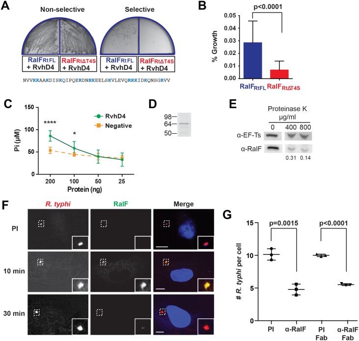
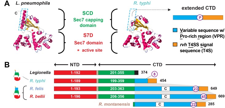
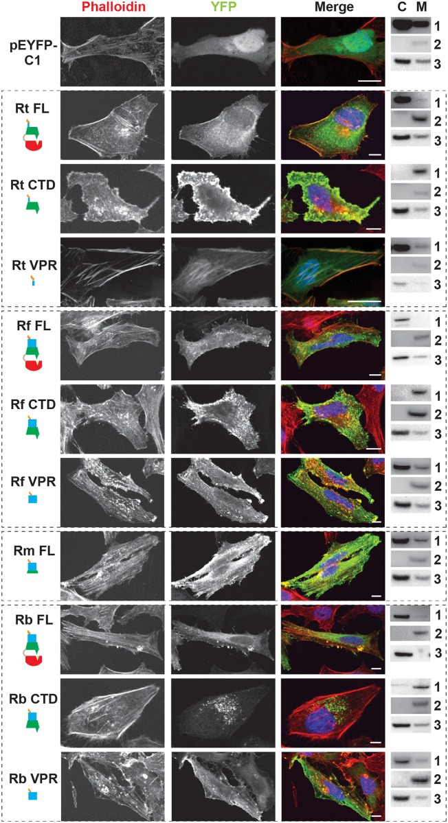
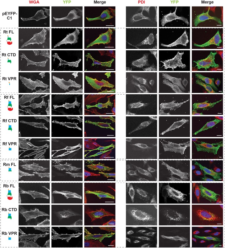
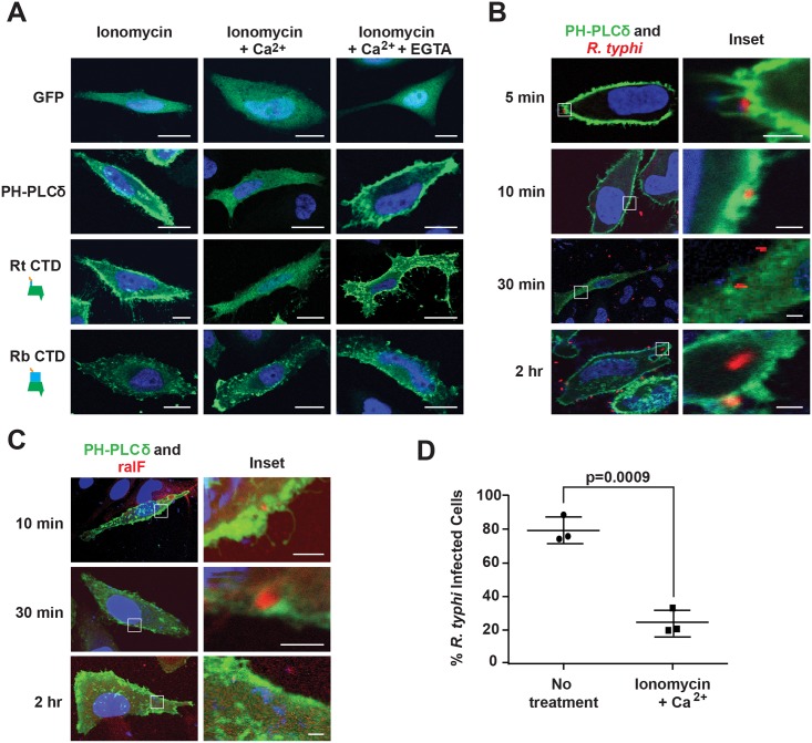
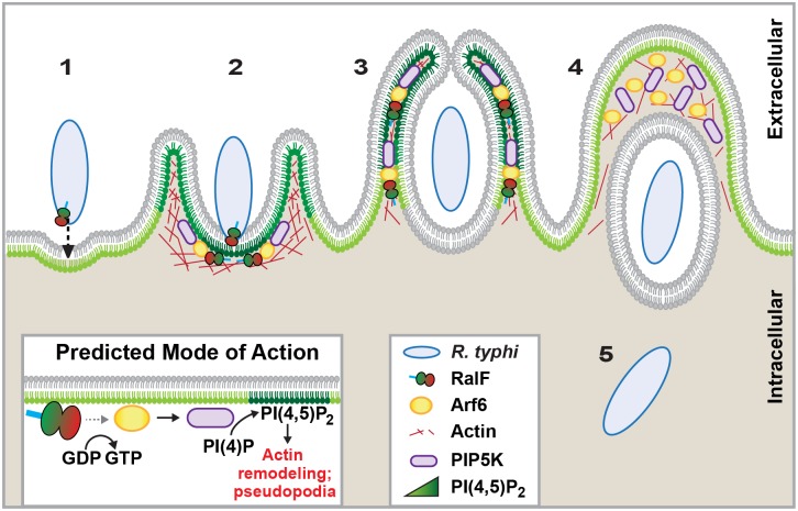
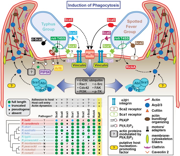
Bacterial Sec7-domain-containing proteins (RalF) are known only from species of Legionella and Rickettsia, which have facultative and obligate intracellular lifestyles, respectively. L. pneumophila RalF, a type IV secretion system (T4SS) effector, is a guanine nucleotide exchange factor (GEF) of ADP-ribosylation factors (Arfs), activating and recruiting host Arf1 to the Legionella-containing vacuole. In contrast, previous in vitro studies showed R. prowazekii (Typhus Group) RalF is a functional Arf-GEF that localizes to the host plasma membrane and interacts with the actin cytoskeleton via a unique C-terminal domain. As RalF is differentially encoded across Rickettsia species (e.g., pseudogenized in all Spotted Fever Group species), it may function in lineage-specific biology and pathogenicity. Herein, we demonstrate RalF of R. typhi (Typhus Group) interacts with the Rickettsia T4SS coupling protein (RvhD4) via its proximal C-terminal sequence. RalF is expressed early during infection, with its inactivation via antibody blocking significantly reducing R. typhi host cell invasion. For R. typhi and R. felis (Transitional Group), RalF ectopic expression revealed subcellular localization with the host plasma membrane and actin cytoskeleton. Remarkably, R. bellii (Ancestral Group) RalF showed perinuclear localization reminiscent of ectopically expressed Legionella RalF, for which it shares several structural features. For R. typhi, RalF co-localization with Arf6 and PI(4,5)P2 at entry foci on the host plasma membrane was determined to be critical for invasion. Thus, we propose recruitment of PI(4,5)P2 at entry foci, mediated by RalF activation of Arf6, initiates actin remodeling and ultimately facilitates bacterial invasion. Collectively, our characterization of RalF as an invasin suggests that, despite carrying a similar Arf-GEF unknown from other bacteria, different intracellular lifestyles across Rickettsia and Legionella species have driven divergent roles for RalF during infection. Furthermore, our identification of lineage-specific Arf-GEF utilization across some rickettsial species illustrates different pathogenicity factors that define diverse agents of rickettsial diseases.


Conflict of interest statement

The authors have declared that no competing interests exist.

Figures

Fig 1
Fig 1. R. typhi RalFRt interacts with RvhD4 and is expressed early during host cell invasion.
(A) Bacterial two-hybrid (B2H) assay reveals an interaction between RalFRt and RvhD4. ralF RtFL and ralF RtΔT4S were cloned into pTRG (prey) and rvhD4 was cloned into pBT (bait) of the B2H system. Constructed bait and prey plasmids were co-transformed into BacterioMatch II reporter electro-competent cells. Transformants were screened on non-selective plate (left) and positive interactions were identified on dual selective screening plate (right). The amino acid sequence deleted from ralF RtΔT4S (positively charged residues are colored blue) is shown at bottom. (B) Quantification of bacterial growth in the B2H assay described in panel A. Percent growth of CFUs of reporter cells harboring recombinant plasmids on dual selective screening medium was calculated relative to CFUs obtained on non-selective medium. Error bars represent mean ± SD of three independent experiments (Student’s two-sided t-test). (C) R. typhi RvhD4 exhibits ATPase activity. A series dilution of purified RvhD4 in assay buffer was incubated with reagent for 30 min at 21°C. The inorganic phosphate (Pi) released from ATP was quantified by measuring absorbance at OD 620 nm. As a negative control, a non-related R. typhi protein (RT0600) was assayed. Error bars represent mean ± SD of three independent experiments. * p = 0.01, **** p<0.0001; Student’s two-sided t-test. (D) Protein immunoblot of recombinant RvhD4 (~64 kDa) used in ATPase activity assays described in panel C. (E) RalFRt is surface exposed. Purified R. typhi was treated with 400 μg/mL or 800 μg/mL Proteinase K or in buffer alone for 1 hr. Lysates were resolved and immunoblotted for RalF or the R. typhi cytoplasmic control protein, elongation factor Ts (EF-Ts). Densitometry was performed using ImageJ and the intensity of RalF was normalized to EF-Ts. Representative image from two independent experiments is shown. Intensity of RalF normalized to EF-Ts and relative to untreated control is shown below the immunoblots. (F) RalF is expressed during early infection. HeLa cells infected with R. typhi for 10 and 30 min were fixed and R. typhi and RalF detected with rat anti-R. typhi (red) and affinity purified rabbit anti-RalFRt (green) antibodies, respectively. DAPI (blue) is shown in the merged image. Boxed regions are enlarged to show detail. Pre-immune (PI) cells were treated with rabbit PI serum in place of anti-RalFRt antibody. (Scale bar: 10 μm). (G) Anti-RalFRt IgG and Fab fragments inhibit R. typhi host cell infection. HeLa cells were infected with partially purified R. typhi pre-absorbed for 30 min with 20μg PI IgG serum, anti-RalFRt IgG, PI Fab fragments or anti-RalFRt Fab fragments. Cells were fixed 2 hrs post infection and R. typhi and the cell membrane detected with anti-R. typhi serum and Alexa Fluor 594 wheat germ agglutinin, respectively. The number of R. typhi per host cell was counted for 100 individual host cells in three independent experiments and normalized to PI serum. Error bars represent mean ± SD (Student’s two-sided t-test).
Fig 2
Fig 2. Characteristics and comparative analysis of bacterial Sec7 domain-containing proteins (RalF).
(A) Comparison of the crystal structure of Legionella pneumophila RalF (PDB 4C7P) [53] with the predicted structure of R. typhi RalF (RT0362). Modeling done with Phyre2 [69]. The delineation of the Sec7 domain (S7D, red) and Sec7-capping domain (SCD, green) is shown, with an approximation of the active site Glu (asterisk), which is essential for Arf recruitment to the Legionella containing vacuole [52]. The distinguishing feature of the otherwise highly similar proteins is the extended C-terminal domain in R. typhi RalF relative to L. pneumophila RalF. The blue dashed box depicts the extended C-terminal domain of Rickettsia RalF sequences, which can be delineated into a variable sequence with Pro-rich region (VPR) and an rvh T4SS signal sequence (T4S). (B) Domain organization of Legionella and Rickettsia RalF proteins. The structural conservation witnessed in panel A is encoded by conserved S7D (S2B Fig) and SCD (S3B Fig) sequences (~45% ID across Legionella and Rickettsia). Rickettsia RalF VPRs vary extensively across homologs; some Rickettsia RalF proteins contain only the VPR and T4S (S4A Fig). C, coiled-coil. Number of Pro residues within purple circles. NCBI GenBank accession numbers for all proteins are provided in the legend of S2 Fig.
Fig 3
Fig 3. RalF subcellular localization and actin filament disruption mediated by the SCD and VPR.
HeLa cells transfected with YFP tagged constructs (green, described in Fig 2B) were stained with Alexa Fluor 594 phalloidin to detect actin (red). DAPI (blue) is shown in the merged image. Cytoplasmic (C) and membrane (M) localization was confirmed via membrane fractionation of HEK293T cells Lipofectamine 2000 transfected with the indicated plasmids followed by immunoblotting. Immunoblot primary antibodies: 1, rabbit anti-GFP (Life Technologies); 2, membrane marker rabbit anti-Calnexin (Abcam); 3, cytoplasmic marker mouse anti-GAPDH (Abcam). Rt, R. typhi; Rf, R. felis; Rm, R. montanensis; Rb, R. bellii. (Scale bar: 10 μm).
Fig 4
Fig 4. Subcellular localization of rickettsial RalF proteins to host membranes.
HeLa cells expressing YFP tagged RalF proteins (green, described in Fig 2) were fixed and stained with Alexa Fluor 594 wheat germ agglutinin (WGA) to detect the plasma membrane (left) or anti-PDI antibody to detect the endoplasmic reticulum (right). DAPI (blue) is shown in the merged image. (Scale bar: 10 μm).
Fig 5
Fig 5. PI(4,5)P2 interacts with RalFRt and mediates R. typhi infection.
(A) RalFRtCTD co-localizes with PI(4,5)P2. HeLa cells transfected with pEYFP-C1 empty vector, GFP-C1-PLCδ-PH (a PI(4,5)P2 biosensor), EYFP–RalFRtCTD, or EYFP–RalFRbCTD were treated with 5 μM ionomycin alone, with Ca2+, or with Ca2+ and EGTA. Nuclei were stained with DAPI (blue). (Scale bar: 10 μm). (B) PI(4,5)P2 is recruited during R. typhi infection. HeLa cells transfected with GFP-C1-PLCδ-PH (green) were infected with R. typhi (MOI ~100:1) for indicated times. R. typhi was detected with rat anti-R. typhi serum and Alexa Fluor 594 anti-rat antibody (red). Nuclei were stained with DAPI (blue). Boxed regions are enlarged to show detail (inset). (Scale bar: 1 μm). (C) RalF localizes to PI(4,5)P2-enriched regions of the plasma membrane during R. typhi infection. HeLa cells transfected with GFP-C1-PLCδ-PH (green) were infected with R. typhi (MOI ~100:1) for indicated times. RalFRt was detected with rabbit anti-RalFRt and Alexa Fluor 594 anti-rabbit antibodies (red). Nuclei were stained with DAPI (blue). Boxed regions are enlarged to show detail (inset). (Scale bar: 1 μm). (D) Ionomycin and Ca2+ treatment decreases R. typhi infection. HeLa cells treated with 5 μM ionomycin and Ca2+ or no treatment were infected with R. typhi (MOI ~100:1) for 2 hrs. R. typhi was detected with rat anti-R. typhi serum and Alexa Fluor 488 anti-rat antibody. Cell membrane was stained with Alexa Fluor 594 wheat germ agglutinin. The number of infected host cells was counted, with percent infection of three independent experiments (100 host cells counted for each) plotted. Error bars represent mean ± SD (Student’s two-sided t-test).
Fig 6
Fig 6. Arf6 is recruited by R. typhi RalF and is required for infection.
(A) Ectopically expressed RalFRtFL co-localizes with Arf6 but not Arf5. HeLa cells co-expressing EYFP, EYFP-RalFRtFL or EYFP-RalFRbFL and mRFP-Arf6 (left) or -Arf5 (right) were fixed with 4% para-formaldehyde. Nuclei were stained with DAPI (blue). (Scale bar: 10 μm). (B) RalFRtFL pull-down of Arf6. Lysates from HEK293T cells expressing mRFP-Arf5 or -Arf6 were incubated with HisPur Cobalt resin bound with rHis-RalFRtFL or resin alone. Bound proteins were eluted with imidazole and analyzed by protein immunoblot using antibodies as indicated. (C) Arf6 is recruited during R. typhi entry. HeLa cells expressing mRFP-Arf5 or -Arf6 (red) were infected with partially purified R. typhi (MOI ~100). Ten minutes post infection, cells were fixed and R. typhi detected with anti-R. typhi serum (green). DAPI (blue) is shown in the merged image. Boxed regions are enlarged to show detail. White arrowheads indicate R. typhi. (Scale bar: 5 μm). (D) Arf6 knockdown inhibits R. typhi infection. HeLa cells transfected with negative, Arf6, or Arf5 siRNA were infected with partially purified R. typhi (MOI ~100). At 2 hrs post infection, cells were fixed, plasma membrane stained with Alexa Fluor 594 wheat germ agglutinin, and R. typhi detected with rat anti-R. typhi serum and Alexa Fluor 488 anti-rat antibody. The number of R. typhi per host cell was counted for 100 host cells for three independent experiments. Error bars represent mean ± SD (Student’s two-sided t-test). (Scale bar: 5μm). (E) Confirmation of Arf6 and Arf5 knockdown. Arf6 and Arf5 knockdown, 80% and 96% respectively, was confirmed by western blot and densitometry analysis using ImageJ (NIH).
Fig 7
Fig 7. Schematic of R. typhi entry.
R. typhi entry has been broken down into five conceptual stages: binding (1); extension of pseudopodia (2); membrane fusion and internalization (3); formation of early endosome (4); bacterial escape from endosome (5). Schematic is a representation of micrographs from Figs 5B, 5C and 6C. Inset depicts hypothetical recruitment and activation of PIP5K via RalFRt-activated Arf6, which results in PI(4,5)P2 enrichment and actin rearrangement to facilitate for R. typhi entry.
Fig 8
Fig 8. Model for the variable pathways utilized by divergent Rickettsia species for host cell entry.
General pathways for Typhus Group (TG, left) and Spotted Fever Group (SFG, right) rickettsiae species are inferred primarily from previous work on SFG rickettsiae species R. conorii [26] and R. parkeri [108] or data from the present study (R. typhi). At center, a conserved proximal hub of the pathway commences with Sca5 binding to host receptor Ku70 [110], which triggers a host-signaling cascade (gray box) involving c-Cbl-mediated ubiquitination of Ku70, Rho-family GTPases Cdc42 and Rac1, phosphoinositide 3-kinase (PI3K) activity, and activation of tyrosine kinases (e.g., c-Src, FAK and p-TK) and their phosphorylated targets. The divergent distal arms of this pathway involve recruitment of factors for activating the actin nucleating complex (Arp2/3), which leads to host actin polymerization, extensive membrane ruffling and filopodia formation, and bacterial internalization in a clathrin and calveolin dependent process. For SFG rickettsiae, the WAVE complex recruits Arp2/3, with its activation via an unknown nucleation promoting factor (either host or bacterial; e.g., RickA). While these processes remain to be characterized for TG rickettsiae, our work suggests that secreted RalF recruits the GTPase Arf6, precipitating an accumulation of PI(4,5)P2 that modulates the activities of a range of actin-associated host proteins (green star). Additional bacterial proteins, some of which are known to facilitate host cell entry, have white lettering with colored boxed backgrounds. Known pathways for protein secretion and host cell receptor-binding, as recently reviewed [45], are shown with solid black lines; all other modeled pathways (shown with dashed lines) are either inferred by homology (e.g., Sca1 of TG rickettsiae as an adhesin based on characterization for Sca1 of R. conorii [36]) or estimated based on in silico analyses (e.g., Sca3 of TG rickettsiae as a putative analog to the α2β1 integrin-binding Sca0 of R. conorii [35]). A phylogenomics analysis across select Rickettsia species (bottom, left) illustrates the genomic variation underlying all of the bacterial components of the models. Adapted from our recent report on the Rickettsia secretome [45]. Red, ancestral group (AG); blue, transitional group (TRG); aquamarine, TG; brown, SFG.

References

    1. Casadevall A. Evolution of intracellular pathogens. Annu Rev Microbiol. 2008;62: 19–33. 10.1146/annurev.micro.61.080706.093305 - DOI - PubMed
    1. Hybiske K, Stephens RS. Exit strategies of intracellular pathogens. Nat Rev Microbiol. 2008;6: 99–110. 10.1038/nrmicro1821 - DOI - PubMed
    1. Kumar Y, Valdivia RH. Leading a sheltered life: intracellular pathogens and maintenance of vacuolar compartments. Cell Host Microbe. 2009;5: 593–601. 10.1016/j.chom.2009.05.014 - DOI - PMC - PubMed
    1. Creasey EA, Isberg RR. Maintenance of vacuole integrity by bacterial pathogens. Current Opinion in Microbiology. 2014. pp. 46–52. 10.1016/j.mib.2013.11.005 - DOI - PMC - PubMed
    1. Garcia-del Portillo F, Finlay BB. The varied lifestyles of intracellular pathogens within eukaryotic vacuolar compartments. Trends Microbiol. 1995;3: 373–380. - PubMed

Publication types

MeSH terms

Substances