. 2015 Oct 5;54(41):11983-8.
doi: 10.1002/anie.201504902. Epub 2015 Aug 20.

Induction of stem-cell-derived functional neurons by NanoScript-based gene repression

Affiliations

Induction of stem-cell-derived functional neurons by NanoScript-based gene repression

Sahishnu Patel et al. Angew Chem Int Ed Engl. .

Abstract

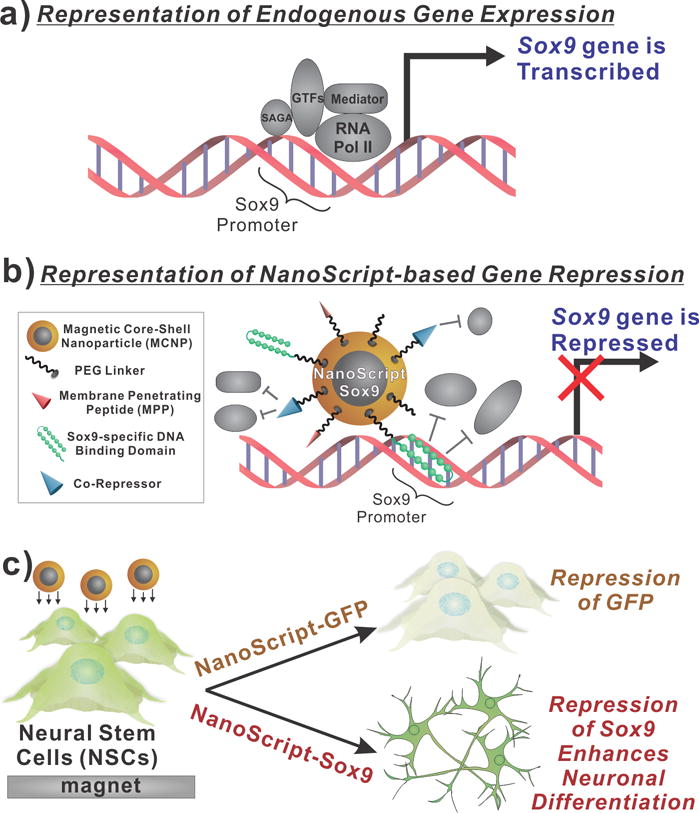
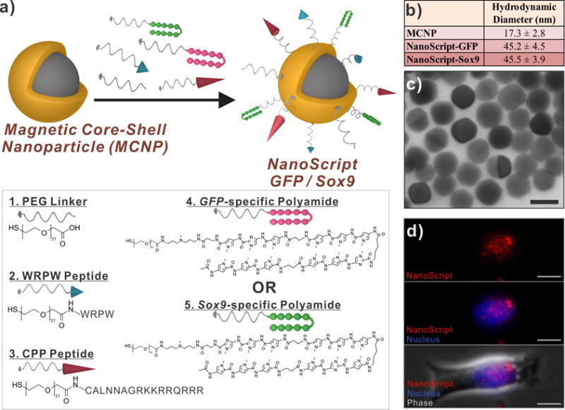
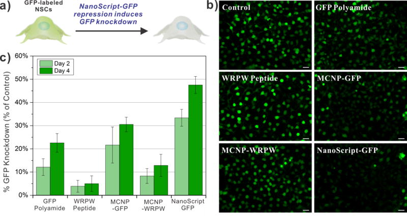
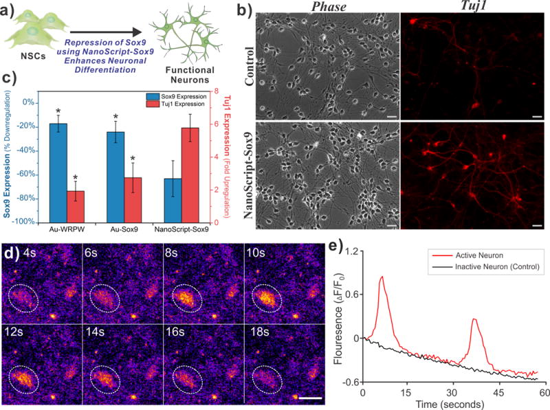
Even though gene repression is a powerful approach to exogenously regulate cellular behavior, developing a platform to effectively repress targeted genes, especially for stem-cell applications, remains elusive. Herein, we introduce a nanomaterial-based platform that is capable of mimicking the function of transcription repressor proteins to downregulate gene expression at the transcriptional level for enhancing stem-cell differentiation. We developed the "NanoScript" platform by integrating multiple gene repression molecules with a nanoparticle. First, we show a proof-of-concept demonstration using a GFP-specific NanoScript to knockdown GFP expression in neural stem cells (NSCs-GFP). Then, we show that a Sox9-specific NanoScript can repress Sox9 expression to initiate enhanced differentiation of NSCs into functional neurons. Overall, the tunable properties and gene-knockdown capabilities of NanoScript enables its utilization for gene-repression applications in stem cell biology.

Keywords: NanoScript; gene knockdown; nanoparticles; neuronal differentiation; transcription repressor proteins.


Figures

Figure 1
Figure 1. Schematic Representation of NanoScript-based Gene Repression
(a) When components of the transcriptional basal complex assemble on a target DNA sequence, such as the Sox9 promoter sequence, the corresponding gene is transcribed. (b) NanoScript-based gene expression is based on the synergistic effect of the DNA Binding Domain molecule for steric hindrance and the co-repressor molecule to disrupt the formation of the transcriptional basal complex on the target DNA sequence. (c) To demonstrate NanoScript-based repression in neural stem cells (NSCs), a GFP-specific NanoScript silences expression of GFP and a Sox9-specific NanoScript represses Sox9 to induce neuronal differentiation.
Figure 2
Figure 2. Construction and Characterization of NanoScript
(a) The magnetic core-shell nanoparticle (MCNP) was functionalized with PEG-terminated biomolecules, through thiol-gold interactions, to develop the NanoScript platform specific for either GFP or Sox9. (b) The hydrodynamic diameter and (c) transmission electron micrographs of NanoScript (scale bar = 20 nm). (d) A dye-labeled NanoScript (red) was transfected into rat NSCs and NanoScript was detected within the nucleus (blue) (scale bar = 20 μm).
Figure 3
Figure 3. NanoScript-GFP Effectively Silences GFP Expression
(a) Schematic representation of NanoScript-GFP downregulating GFP expression (i.e. reduction in green fluorescence from the cell) in GFP-labeled rat NSCs. (b) Fluorescence images of GFP-labeled rNSCs 4 days post transfection showing a maximal decrease in GFP expression (green) when NanoScript-GFP is transfected (scale bar = 20 μm). A 1 nM concentration of MCNPs was applied during the transfection. (c) Quantification of GFP expression in the fluorescence images corroborates the trend in the images and reveals that NanoScript-GFP has the highest GFP knockdown as compared to the controls. Quantification of GFP knockdown is an average from 6 images and standard error is from three independent trials.
Figure 4
Figure 4. NanoScript-Sox9 Represses Sox9 to Induce Functional Neuronal Differentiation
(a) Schematic representation of Sox9 repression in human NSCs by NanoScript-Sox9 induces enhanced neuronal differentiation. (b) Fluorescence images of hNSC stained with Tuj1 5 days post-transfection shows greater Tuj1 expression (red) when NanoScript-Sox9 is transfected. (Scale bar = 20 μm). (c) Gene expression analysis using qPCR in hNSCs reveals that repression of Sox9 correlates with an upregualtion of Tuj1. (Percent down-regulation of Sox9 and fold up-regulation of Tuj1 was calculated by normalizing to the housekeep gene, GAPDH, from the control) Standard error is from three independent trials (* = P < 0.05). (d) Spontaneous calcium fluctuations via Fluo4 fluorescence (orange/yellow color) for an active neuron (white circle) during 18 seconds of imaging (scale bar = 20 μm). (e) Traces for the normalized fluorescence change (ΔF/F0) representing spontaneous calcium ion influx for an active neuron (red line) and an inactive neuron (black line). Decreasing trend of the fluorescence is due to mild photobleaching.

References

    1. Guenther MG, Frampton GM, Soldner F, Hockemeyer D, Mitalipova M, Jaenisch R, Young RA. Cell Stem Cell. 2010;7:249–257. - PMC - PubMed
    1. Rockman MV, Kruglyak L. Nat Rev Genet. 2006;7:862–872. - PubMed
    1. Reik W. Nature. 2007;447:425–432. - PubMed
    1. Spitz F, Furlong EE. Nat Rev Genet. 2012;13:613–626. - PubMed
    1. Weake VM, Workman JL. Nat Rev Genet. 2010;11:426–437. - PubMed
    2. Mapp AK, Ansari AZ, Ptashne M, Dervan PB. Proc Natl Acad Sci U S A. 2000;97:3930–3935. - PMC - PubMed

Publication types

MeSH terms

LinkOut - more resources