Clinical Trial
. 2015 Aug 20;10(8):e0136109.
doi: 10.1371/journal.pone.0136109. eCollection 2015.

Discovery of Novel Plasmodium falciparum Pre-Erythrocytic Antigens for Vaccine Development

Affiliations
Clinical Trial

Discovery of Novel Plasmodium falciparum Pre-Erythrocytic Antigens for Vaccine Development

Joao C Aguiar et al. PLoS One. .

Abstract

Background: Nearly 100% protection against malaria infection can be achieved in humans by immunization with P. falciparum radiation-attenuated sporozoites (RAS). Although it is thought that protection is mediated by T cell and antibody responses, only a few of the many pre-erythrocytic (sporozoite and liver stage) antigens that are targeted by these responses have been identified.

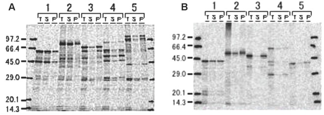
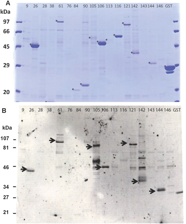
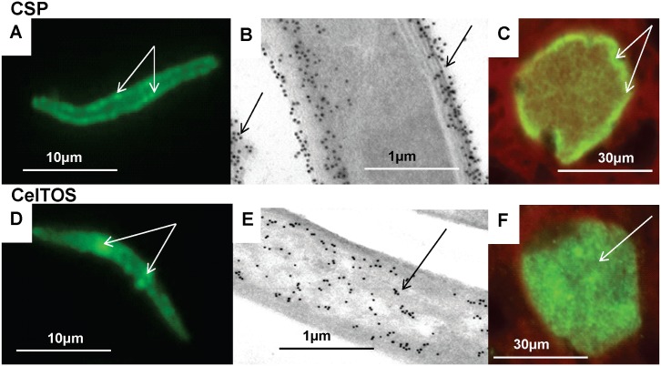
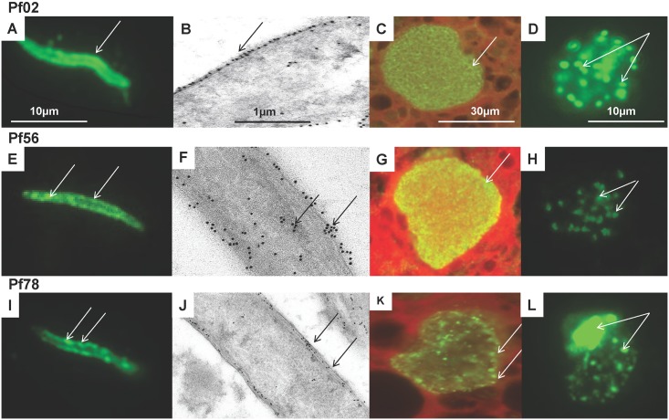
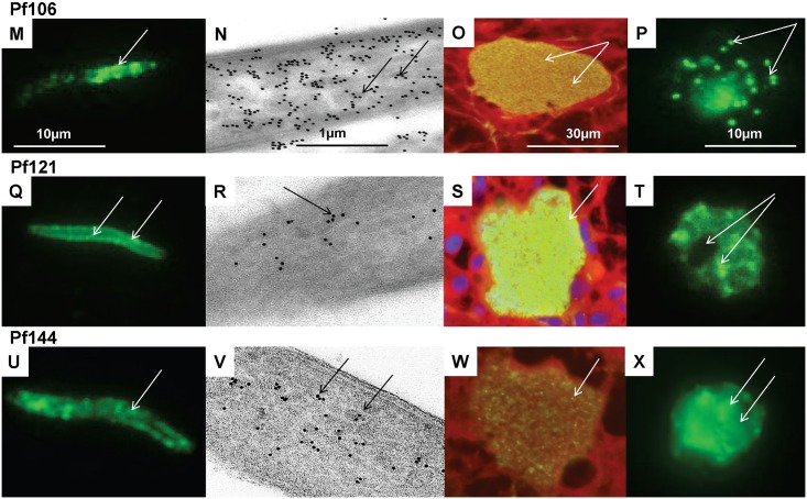
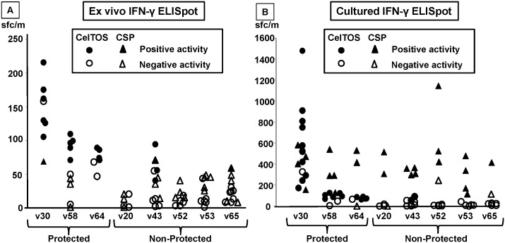
Methodology: Twenty seven P. falciparum pre-erythrocytic antigens were selected using bioinformatics analysis and expression databases and were expressed in a wheat germ cell-free protein expression system. Recombinant proteins were recognized by plasma from RAS-immunized subjects, and 21 induced detectable antibody responses in mice and rabbit and sera from these immunized animals were used to characterize these antigens. All 21 proteins localized to the sporozoite: five localized to the surface, seven localized to the micronemes, cytoplasm, endoplasmic reticulum or nucleus, two localized to the surface and cytoplasm, and seven remain undetermined. PBMC from RAS-immunized volunteers elicited positive ex vivo or cultured ELISpot responses against peptides from 20 of the 21 antigens.

Conclusions: These T cell and antibody responses support our approach of using reagents from RAS-immunized subjects to screen potential vaccine antigens, and have led to the identification of a panel of novel P. falciparum antigens. These results provide evidence to further evaluate these antigens as vaccine candidates.

Trial registration: ClinicalTrials.gov NCT00870987 ClinicalTrials.gov NCT00392015.


Conflict of interest statement

Competing Interests: JCA is an employee of Camris International. JW is an employee of Technical Resources International, Inc. JKM is an employee of EMD Millipore Corporation. There are no patents, products in development, or marketed products to declare. This does not alter the authors' adherence to all the PLOS ONE policies on sharing data and materials.

Figures

Fig 1
Fig 1. High-throughput expression and detection of P. falciparum proteins.
Compartmental wheat germ expression of recombinant proteins represented by five clones as GST (A) and 6xHis fusions (B). [C14]-Leucine labeled proteins detected by radio blot in three fractions; total (T), supernatant (S), and pellet (P). Protein predicted molecular weight sizes are: (1) 32.8 kDa, (2) 50.2 kDa, (3) 37.7 kDa, (4) 59.7 kDa and (5) 32.5 kDa. The molecular sizes for GST-fused proteins (A) include an additional 29 kDa (GST molecular size). The autoradiographs for all the 151 clones are shown in S1 Fig.
Fig 2
Fig 2. Expression and detection of affinity purified P. falciparum proteins.
(A) Coomassie blue stained SDS-PAGE gels of 17 randomly-selected GST-fusion proteins that were affinity purified and cleaved. Asterisk indicates purified protein. (B) Western blot probed with pooled RAS-immune sera. Arrow indicates positive reactivity.
Fig 3
Fig 3. Stage-specific expression of CSP and CelTOS by immunofluorescence IFA and immuno-electron microscopy.
CSP and CelTOS were localized to sporozoites (A, B, D, and E) and 7-day old liver stages (C and F) by IFA (A, C, D, F) and by immune-electron microscopy (B, E). CSP: localized to the sporozoite surface, (arrows, A), outer sporozoites membranes and shed material (arrows B), and the periphery of 5 day liver stage (arrows, C). CelTOS: localized to patches within sporozoites (arrows, D) that are associated with micronemes (arrows, E), was not on the surface, and was in the cytoplasm of five day liver stages (arrow, F).
Fig 4
Fig 4. Stage-specific expression of three novel P. falciparum antigens by immunofluorescence and immune-electron microscopy.
Three novel P. falciparum antigens (Pf02, Pf56, Pf78) were localized by immunofluorescence to sporozoites (A, E, I), 7-day liver stages (C, G, K), and blood stages (D, H, L); and by immuno-electron microscopy to sporozoites (B, F, J). Pf02: localized to the sporozoite surface (A), specifically the outer membrane (arrow, B), the cytoplasm of liver stages (C) and cytoplasm of blood stage merozoites (arrows, D). Pf56: localized to patches in sporozoite periphery (arrows, E, F) and sporozoite interior (arrows, F), the cytoplasm of liver stages (arrow, G) and cytoplasm of blood stage merozoites (arrows, H). Pf78: localized as discrete clumps on the sporozoite periphery (arrows, I), that were mostly in the middle and inner surface membranes (arrows, J), as discrete clumps in the liver stage periphery (arrows, K), and in the blood stage schizonts residual body and cytoplasm, but not merozoites (arrows, L).
Fig 5
Fig 5. Stage-specific expression of three novel P. falciparum antigens by immunofluorescence and immune-electron microscopy.
Three additional novel P. falciparum antigens (Pf106, Pf121, Pf144) were localized by immunofluorescence to sporozoites (M, Q, U), 7-day liver stages (O, S, W), and blood stages (P, T, X); and by immuno-electron microscopy to sporozoites (N, R, V). Pf106: localized in clumps at the sporozoite anterior pole (arrow, M), in vesicles inside the sporozoite (arrows, N), in liver stage merozoites (arrows, 0), and in blood stage merozoites (arrows, P). Pf121: localized unevenly in the sporozoite periphery/interior (arrows, Q) that appear to localize to micronemes (arrows, R), throughout liver stage parasite (arrow, S) and in clumps in blood stage schizonts but not the residual body (arrows, T). Pf144: localized to clumps inside the sporozoite (arrow, U) that are associated with micronemes (arrows, V), but is only weakly detected in 7-day liver stage parasites (arrow, W) and is dispersed in the blood stage schizonts (arrow, X).
Fig 6
Fig 6. Antigen-specific IFN-γ ELISpot activity of protected and non-protected RAS-immunized volunteers targeting CelTOS and CSP.
CelTOS (circles) and CSP (triangles) overlapping peptide pools spanning full length CSP or CelTOS were used to stimulate T cell responses of three protected (v30, v58, v64) and five non-protected (v20, v43, v52, v53, v65) RAS-immunized subjects, and responses were measured by IFN-γ ELISpot, and expressed as spot-forming cells/million PBMC (sf/m). A. In ex vivo assays, samples were scored as positive (filled markers) using high stringency criteria (significant difference between test antigen and medium controls using Student’s t-test, at least a doubling and a difference of at least 10 sfc/m (See Methods) or as negative (open markers). Occasional values of activities that were negative exceeded positive values if the pre-immunization activities were high. Overall, the CelTOS-specific T cell responses among protected subjects (geometric mean 98 sfc/m) were significantly higher than the CelTOS-specific T cell responses of non-protected subjects (geometric mean 12 sfc/m, p = <0.001, Mann-Whitney U test), whereas CSP-specific T cell responses were similar among protected (geometric mean 18 sfc/m) and non-protected (geometric mean 11 sfc/m, not significant) subjects. B. In cultured ELISpot assays, CelTOS-specific T cell responses of protected subjects (geometric mean 505 sfc/m) were also significantly higher than non-protected subjects (geometric mean 157 sfc/m, p = 0.002, Mann-Whitney U test). CSP-specific T cell responses of protected subjects (geometric mean 215 sfc/m) were similar to those of non-protected subjects (geometric mean 235 sfc/m, not significant).

References

    1. Weiss WR, Good MF, Hollingdale MR, Miller LH, Berzofsky JA. Genetic control of immunity to Plasmodium yoelii sporozoites. J Immunol. 1989;143(12):4263–6. - PubMed
    1. Nussenzweig V, Nussenzweig RS. Circumsporozoite proteins of malaria parasites. Cell. 1985;42(2):401–3. - PubMed
    1. Gwadz RW, Cochrane AH, Nussenzweig V, Nussenzweig RS. Preliminary studies on vaccination of rhesus monkeys with irradiated sporozoites of Plasmodium knowlesi and characterization of surface antigens of these parasites. Bull World Health Organ. 1979;57 Suppl 1:165–73. - PMC - PubMed
    1. Clyde DF. Immunization of man against falciparum and vivax malaria by use of attenuated sporozoites. Am J Trop Med Hyg. 1975;24(3):397–401. - PubMed
    1. Rieckmann KH, Beaudoin RL, Cassells JS, Sell KW. Use of attenuated sporozoites in the immunization of human volunteers against falciparum malaria. Bull World Health Organ. 1979;57 Suppl 1:261–5. - PMC - PubMed

Publication types

MeSH terms

Associated data