. 2015 Aug 21:5:13412.
doi: 10.1038/srep13412.

Sonorensin: A new bacteriocin with potential of an anti-biofilm agent and a food biopreservative

Affiliations

Sonorensin: A new bacteriocin with potential of an anti-biofilm agent and a food biopreservative

Lipsy Chopra et al. Sci Rep. .

Abstract

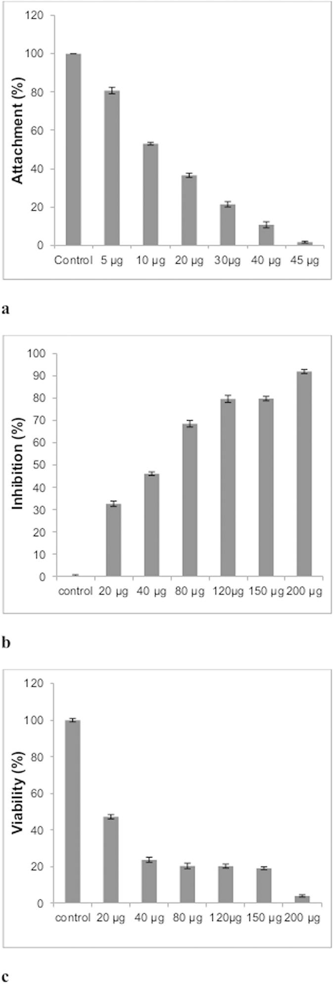
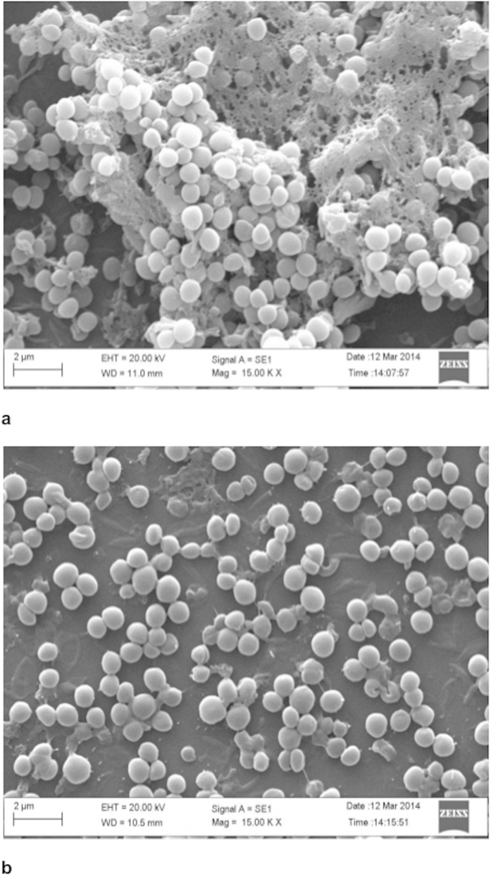
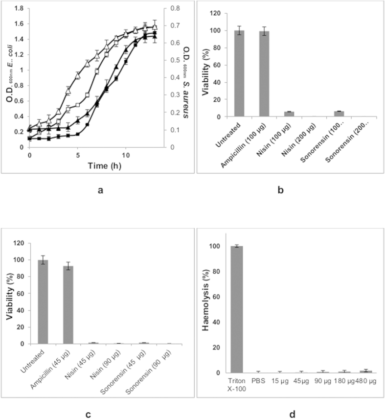
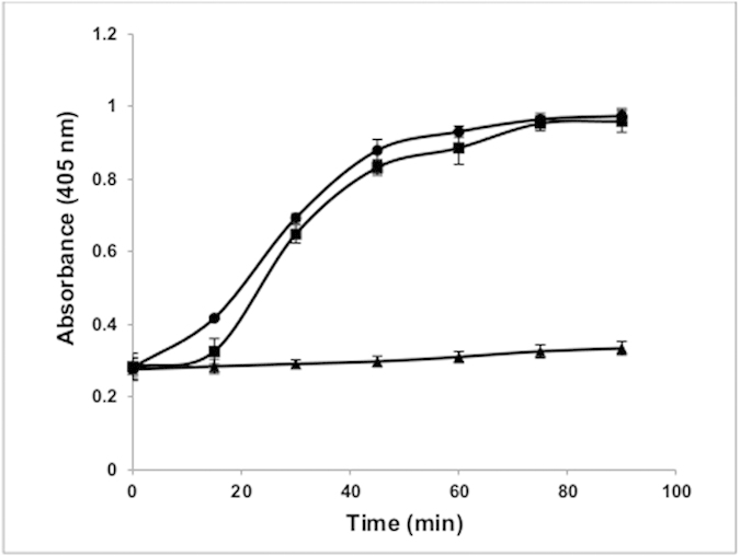
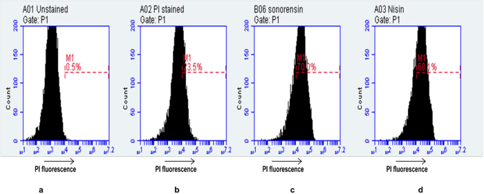
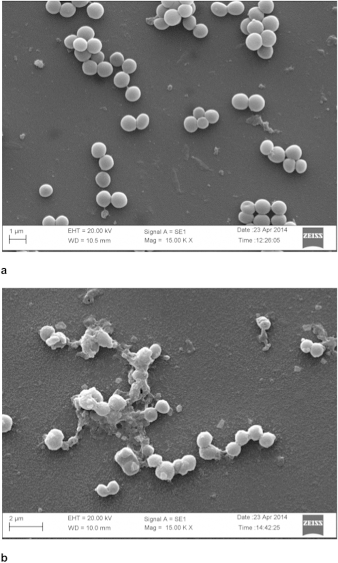
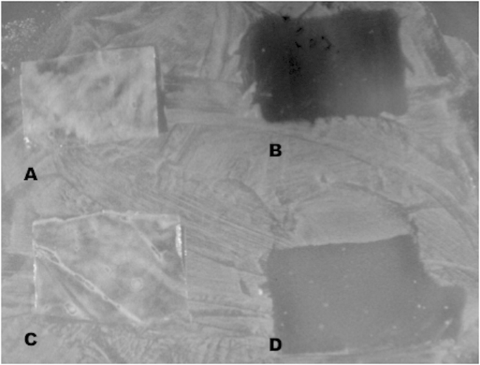
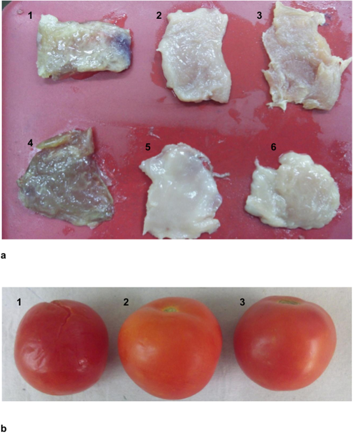
The emergence of antibiotic resistant bacteria has led to exploration of alternative therapeutic agents such as ribosomally synthesized bacterial peptides known as bacteriocins. Biofilms, which are microbial communities that cause serious chronic infections, form environments that enhance antimicrobial resistance. Bacteria in biofilm can be upto thousand times more resistant to antibiotics than the same bacteria circulating in a planktonic state. In this study, sonorensin, predicted to belong to the heterocycloanthracin subfamily of bacteriocins, was found to be effectively killing active and non-multiplying cells of both Gram-positive and Gram-negative bacteria. Sonorensin showed marked inhibition activity against biofilm of Staphylococcus aureus. Fluorescence and electron microscopy suggested that growth inhibition occurred because of increased membrane permeability. Low density polyethylene film coated with sonorensin was found to effectively control the growth of food spoilage bacteria like Listeria monocytogenes and S. aureus. The biopreservative effect of sonorensin coated film showing growth inhibition of spoilage bacteria in chicken meat and tomato samples demonstrated the potential of sonorensin as an alternative to current antibiotics/ preservatives.


Figures

Figure 1
Figure 1. Effect of sonorensin on S. aureus biofilms.
(a) Inhibition of attachment of S. aureus cells by sonorensin at various concentrations (b) Inhibition of biofilm formation (c) Viability of S. aureus cells biofilm (assayed by XTT). Control bars indicate S. aureus cells without any treatment taken as 100% in case of a & c and 0% in case of b. Each well of 96 -well plate contains 4 × 106 CFU S. aureus cells and variable concentration of sonorensin in 200 μl of BHI-sucrose. The plates were incubated at 37 °C for 4 h and 24 h for biofilm attachment assay (a) and inhibition of biofilm formation (b), respectively. (a) About 1.8 ± 0.05% attachment of biofilm was observed in the presence of 1X MIC of sonorensin. (b) Sonorensin showed significant inhibitory activity against S. aureus biofilm formation at 24 h, (c) Reduced XTT conversion was observed in wells with higher sonorensin concentrations and the control (without treatment) showed maximum reduction of XTT. The results were presented as mean ± SD and differences between the control and treated samples were statistically significant (n = 3) (p < 0.005).
Figure 2
Figure 2. The scanning electron micrographs of mature (48 h old) biofilm of S. aureus cells (a) without sonorensin treatment and (b) after sonorensin treatment (50 μg/ml).
The biofilms without sonorensin treatment consisted of nearly uniform, thick layer of cells embedded within a self produced matrix whereas thinning of mature biofilms was observed after sonorensin treatment.
Figure 3
Figure 3. Effect of sonorensin on non-multiplying bacteria.
(a) Comparative growth curves of active (open symbols) and non-multiplying (filled symbols) cells of E. coli (squares) and S. aureus (triangles). Cell viability was used as a measure of the effect of sonorensin on non-multiplying cells of (b) E. coli and (c) S. aureus. The CFU count from the untreated sample was taken as 100% in the cell viability calculations. An extended lag phase of non- multiplying cells compared to their vegetative counterparts (a). The sensitivity to nisin and tolerance to ampicillin confirmed non-multiplying state of cells. Sonorensin was effective against non-multiplying cells of both E. coli (b) and S. aureus (c,d) Sonorensin toxicity to mammalian cells was assayed by measuring its haemolytic activity. Triton X-100 and PBS served as controls. Sonorensin, at concentrations toxic to vegetative and dormant cells of E. coli and S. aureus, had virtually no effect on RBCs and only 1.7 ± 0.04% haemolysis was observed at higher concentrations of sonorensin. All the experiments were repeated three times in triplicate. The results were presented as mean ± SD and differences between the control and treated samples were statistically significant (n = 3) (p < 0.005).
Figure 4
Figure 4. The cytoplasmic membrane permeabilization of S. aureus cells treated with sonorensin (squares) and nisin (circles).
The untreated S. aureus cells (triangles) were taken as control. When the cytoplasmic membrane was permeable ONPG entered the cytoplasm and degraded by β-galactosidase, producing O-nitrophenol that showed absorbance at 405 nm. Sonorensin induced an increase in the permeability of S. aureus. The experiment was carried out three times in triplicate. The results were presented as mean ± SD and differences between the control and treated samples were statistically significant (n = 3) (p < 0.005).
Figure 5
Figure 5. Flow cytometry analysis of effects of sonorensin and nisin on membrane integrity of S. aureus cells.
Data were displayed as flow cytometric histograms of counted bacterial events (y-axis) associated cell fluorescence (x-axis). Marker M1 is the region that the damaged cells were stained by PI. (a) Unstained S. aureus cells (b) Untreated, PI stained S. aureus cells (c) Sonorensin treated, PI stained S. aureus cells, (d) nisin treated, PI stained S. aureus cells. For each sample 104 cells were analysed. The membrane integrity of S. aureus cells was destroyed by treatment with sonorensin.
Figure 6
Figure 6. The scanning electron micrographs of S. aureus cells
(a) without sonorensin treatment, and (b) after sonorensin treatment (50 μg/ml) for 4 h. The treatment of S. aureus with sonorensin displayed roughening of cell surface with cell debris while smooth cell surface was observed in cells without treatment.
Figure 7
Figure 7. Inhibitory activity of coated LDPE films against S. aureus.
(a,c) control; (b) with sonorensin coated LDPE film; (d) with nisin coated LDPE film. The growth of S. aureus was inhibited by sonorensin and nisin coated LDPE films whereas S. aureus grew homogeneously on the surface of the plate and underneath the untreated LDPE film used as control.
Figure 8
Figure 8
Preservative effect of coated LDPE film during the storage of (a) meat (b) tomatoes. (a) Meat samples were spiked with L. monocytogenes (1–3) and S. aureus (4–6). Spoilage of meat is visible in meat samples packaged in control LDPE films (1 & 4) whereas no spoilage was observed in samples packaged with sonorensin (2 & 5) and nisin (3 & 6) coated LDPE films. (b) Tomato sample (1) packaged in untreated LDPE films showed signs of spoilage in contrast to no spoilage in case of tomatoes packaged in sonorensin (2) and nisin (3) coated LDPE films.

References

    1. Monroe D. Looking for chinks in the armor of bacterial biofilms. PLoS Biol. 5, e307 (2007). - PMC - PubMed
    1. Hall-Stoodley L., Costerton J. W. & Stoodley P. Bacterial biofilms: from the natural environment to infectious diseases. Nature Rev. Microbiol. 2, 95–108 (2004). - PubMed
    1. Stewart P. S. & Costerton J. W. Antibiotic resistance of bacteria in biofilms. Lancet 358, 135–138 (2001). - PubMed
    1. Di Lorenzo A. et al. Characterization and performance of a toluene-degrading biofilm developed on pumice stones. Microb. Cell Fact. 4, 4 (2005). - PMC - PubMed
    1. Giaouris E. D. & Nychas G. J. E. The adherence of Salmonella enteritidis PT4 to stainless steel: the importance of the air-liquid interface and nutrient availability. Food Microbiol. 23, 747–752 (2006). - PubMed

MeSH terms