Review
. 2015 Nov;98(5):713-25.
doi: 10.1189/jlb.3RI0615-239R. Epub 2015 Aug 20.

Parainflammation, chronic inflammation, and age-related macular degeneration

Affiliations
Review

Parainflammation, chronic inflammation, and age-related macular degeneration

Mei Chen et al. J Leukoc Biol. 2015 Nov.

Abstract

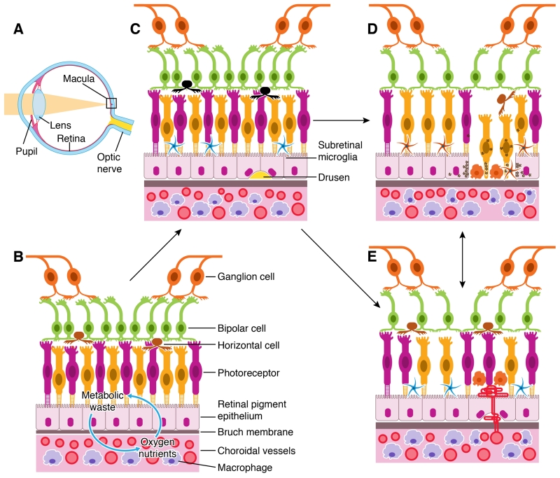
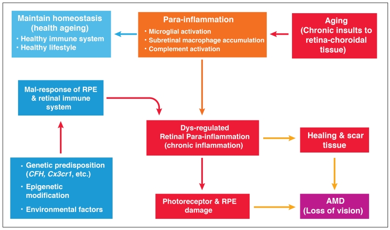
Inflammation is an adaptive response of the immune system to noxious insults to maintain homeostasis and restore functionality. The retina is considered an immune-privileged tissue as a result of its unique anatomic and physiologic properties. During aging, the retina suffers from a low-grade chronic oxidative insult, which sustains for decades and increases in level with advancing age. As a result, the retinal innate-immune system, particularly microglia and the complement system, undergoes low levels of activation (parainflammation). In many cases, this parainflammatory response can maintain homeostasis in the healthy aging eye. However, in patients with age-related macular degeneration, this parainflammatory response becomes dysregulated and contributes to macular damage. Factors contributing to the dysregulation of age-related retinal parainflammation include genetic predisposition, environmental risk factors, and old age. Dysregulated parainflammation (chronic inflammation) in age-related macular degeneration damages the blood retina barrier, resulting in the breach of retinal-immune privilege, leading to the development of retinal lesions. This review discusses the basic principles of retinal innate-immune responses to endogenous chronic insults in normal aging and in age-related macular degeneration and explores the difference between beneficial parainflammation and the detrimental chronic inflammation in the context of age-related macular degeneration.

Keywords: blood-retina barrier; complement; immune privilege; microglia; retina.


Figures

Figure 1
Figure 1. Retina and RPE-choroid in normal, early and late AMD
A, diagram of a human eye. Light pass through the pupil and then focused by lens to the macula of the retinal layer at the back of the eye. B, the retina consists of three layers of neurons, photoreceptor, bipolar and ganglion cells. The RPE monolayer together with Bruch’s membrane (BM) form the outer blood retinal barrier (oBRB) that separates the neuroretina from the choroid. Oxygen and nutrients are transported from the choroid into the outer retina whereas retinal metabolic wastes are transported to choroid through the oBRB. C, the early stages of AMD is characterized by the presence of large Drusen deposits between the RPE and BM, RPE senescence, and the accumulation of microglia and macrophages in the subretinal space. D, geographic atrophy (GA) is typified by the loss of RPE and photoreceptors, accompanied by macrophage infiltration at the lesion site. E, neovascular AMD (nAMD) is caused by the growth of choroidal vessels into the sub-RPE or sub-retina of the macula.
Figure 2
Figure 2. Retinal microglial in health and disease
A, confocal image of a normal mouse retina stained with Iba-1 (green) and Propidium iodide (PI). Iba-1+ microglial cells are located in the ganglion layer (GL), inner plexiform layer (IPL) and outer plexiform layer (OPL). B-C, confocal images taken from the IPL (B) and OPL (C) of a normal mouse eye showing the ramified morphology of resting microglia. D, high-magnification of resting retinal microglia. E, microglia from a 18 month old mouse retina showing heterogeneous activation. Two cells (arrows) demonstrate shorter dendrites (signs of mild activation). F, microglia from paraquat treated mouse eyes demonstrate ameboid shape with a larger cell body, multiple vacuoles and shorter dendrites (signs of full activation). Mildly activated microglia may undergo full activation under acute stress conditions. INL – inner nuclear layer; ONL – outer nuclear layer, RPE – retinal pigment epithelium.
Figure 3
Figure 3. Retinal microglial in the aging eye
A-B, Reconstructed z-stack confocal images from a 16-month (A) and 27-month (B) old mouse retina stained for Iba-1 (green for microglia) and lectin B4 (red for blood vessels). A, at 16 months, few Iba-1+ microglial cells were detected at the subretinal space (short arrows) and some were still connected to cells in the OPL layer (small arrows). B, at 27 months many more Iba-1+ cells were detected at the subretinal space (short arrows), and no Iba-1+ cells were detected between the OPL and subretinal space. C, heterogeneous morphology of Iba-1+ cells at the subretinal space in a 18-month old mouse. Most of the cells have larger cell bodies and shorter dendrites, and a few cells display a relatively small cell body and long dendrites (arrow). D, subretinal Iba-1+ cells from a 27-month old mice showing pigmented cell body (arrowheads). IPL – inner plexiform layer; OPL – outer plexiform layer.
Figure 4
Figure 4. Dysregulated para-inflammation and the pathogenesis of AMD
As we age, oxidative insults accumulate in the macula. A para-inflammatory response characterized by microglial activation, subretinal accumulation and complement activation is initiated to promote macular repair. A healthy immune system should be able to maintain macular homeostasis through para-inflammation. In AMD, the para-inflammatory response is dysregulated due to a) genetic predisposition, b) epigenetic modification, or c) environmental factors. The dysregulated para-inflammation (i.e., chronic inflammation) results in various pro-inflammatory cytokine production or inflammasome activation, which damages RPE and the photoreceptors and leads to the development of AMD. Sustained chronic inflammation at the macula may also lead to scar formation, which can also lead to loss of vision.

References

    1. Mangge H, Almer G, Truschnig-Wilders M, Schmidt A, Gasser R, Fuchs D. Inflammation, adiponectin, obesity and cardiovascular risk. Curr.Med.Chem. 2010;17:4511–4520. - PubMed
    1. Mathieu P, Lemieux I, Despres JP. Obesity, inflammation, and cardiovascular risk. Clin.Pharmacol.Ther. 2010;87:407–416. - PubMed
    1. Lathe R, Sapronova A, Kotelevtsev Y. Atherosclerosis and Alzheimer--diseases with a common cause? Inflammation, oxysterols, vasculature. BMC Geriatr. 2014;14 36-2318-14-36. - PMC - PubMed
    1. Stoll G, Bendszus M. Inflammation and atherosclerosis: novel insights into plaque formation and destabilization. Stroke. 2006;37:1923–1932. - PubMed
    1. McGeer PL, McGeer EG. Inflammation and the degenerative diseases of aging. Ann.N.Y.Acad.Sci. 2004;1035:104–116. - PubMed

Publication types