

Advanced MRI for carotid plaque imaging
- PMID: 26293362
- PMCID: PMC4706840
- DOI: 10.1007/s10554-015-0743-6
Advanced MRI for carotid plaque imaging
Abstract
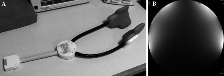
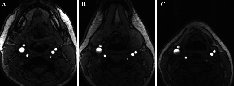
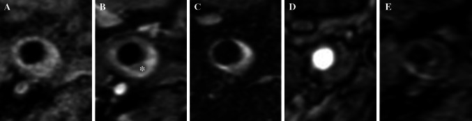
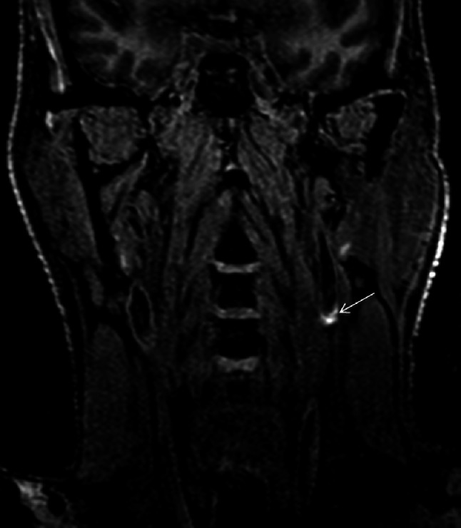
Atherosclerosis is the ubiquitous underling pathological process that manifests in heart attack and stroke, cumulating in the death of one in three North American adults. High-resolution magnetic resonance imaging (MRI) is able to delineate atherosclerotic plaque components and total plaque burden within the carotid arteries. Using dedicated hardware, high resolution images can be obtained. Combining pre- and post-contrast T1, T2, proton-density, and magnetization-prepared rapid acquisition gradient echo weighted fat-saturation imaging, plaque components can be defined. Post-processing software allows for semi- and fully automated quantitative analysis. Imaging correlation with surgical specimens suggests that this technique accurately differentiates plaque features. Total plaque burden and specific plaque components such as a thin fibrous cap, large fatty or necrotic core and intraplaque hemorrhage are accepted markers of neuroischemic events. Given the systemic nature of atherosclerosis, emerging science suggests that the presence of carotid plaque is also an indicator of coronary artery plaque burden, although the preliminary data primarily involves patients with stable coronary disease. While the availability and cost-effectiveness of MRI will ultimately be important determinants of whether carotid MRI is adopted clinically in cardiovascular risk assessment, the high accuracy and reliability of this technique suggests that it has potential as an imaging biomarker of future risk.
Keywords: Atherosclerosis; Cardiovascular risk; Carotid; Imaging; MRI.
Figures

Similar articles
-
Intra-individual comparison of carotid and femoral atherosclerotic plaque features with in vivo MR plaque imaging.Int J Cardiovasc Imaging. 2015 Dec;31(8):1611-8. doi: 10.1007/s10554-015-0737-4. Epub 2015 Aug 22. Int J Cardiovasc Imaging. 2015. PMID: 26296806
-
Ex vivo differential phase contrast and magnetic resonance imaging for characterization of human carotid atherosclerotic plaques.Int J Cardiovasc Imaging. 2015 Oct;31(7):1425-34. doi: 10.1007/s10554-015-0706-y. Epub 2015 Jul 16. Int J Cardiovasc Imaging. 2015. PMID: 26179860
-
In-vivo quantitative T2 mapping of carotid arteries in atherosclerotic patients: segmentation and T2 measurement of plaque components.J Cardiovasc Magn Reson. 2013 Aug 16;15(1):69. doi: 10.1186/1532-429X-15-69. J Cardiovasc Magn Reson. 2013. PMID: 23953780 Free PMC article.
-
Contemporary carotid imaging: from degree of stenosis to plaque vulnerability.J Neurosurg. 2016 Jan;124(1):27-42. doi: 10.3171/2015.1.JNS142452. Epub 2015 Jul 31. J Neurosurg. 2016. PMID: 26230478 Review.
-
Imaging of the high-risk carotid plaque: magnetic resonance imaging.Semin Vasc Surg. 2017 Mar;30(1):54-61. doi: 10.1053/j.semvascsurg.2017.04.009. Epub 2017 Apr 27. Semin Vasc Surg. 2017. PMID: 28818259 Review.
Cited by
-
Predictive values of carotid high-resolution magnetic resonance imaging for large embolus shedding in carotid artery stenting.Turk J Med Sci. 2022 Apr;52(2):286-293. doi: 10.55730/1300-0144.5314. Epub 2022 Apr 14. Turk J Med Sci. 2022. PMID: 36161619 Free PMC article.
-
Cardiovascular risk scoring and magnetic resonance imaging detected subclinical cerebrovascular disease.Eur Heart J Cardiovasc Imaging. 2020 Jun 1;21(6):692-700. doi: 10.1093/ehjci/jez226. Eur Heart J Cardiovasc Imaging. 2020. PMID: 31565735 Free PMC article.
-
Age-Specific Sex Differences in Magnetic Resonance Imaging-Depicted Carotid Intraplaque Hemorrhage.Stroke. 2017 Aug;48(8):2129-2135. doi: 10.1161/STROKEAHA.117.017877. Epub 2017 Jul 13. Stroke. 2017. PMID: 28706117 Free PMC article.
-
Plaque imaging volume analysis: technique and application.Cardiovasc Diagn Ther. 2020 Aug;10(4):1032-1047. doi: 10.21037/cdt.2020.03.01. Cardiovasc Diagn Ther. 2020. PMID: 32968659 Free PMC article. Review.
-
Comparison of four MR carotid surface coils at 3T.PLoS One. 2019 Mar 4;14(3):e0213107. doi: 10.1371/journal.pone.0213107. eCollection 2019. PLoS One. 2019. PMID: 30830934 Free PMC article.
References
Publication types
MeSH terms
Grants and funding
LinkOut - more resources
Full Text Sources
Other Literature Sources
Medical
Miscellaneous
