Comparative Study
. 2015 Oct;35(10):2092-103.
doi: 10.1161/ATVBAHA.115.305843. Epub 2015 Aug 20.

Monocyte-Derived Dendritic Cells Upregulate Extracellular Catabolism of Aggregated Low-Density Lipoprotein on Maturation, Leading to Foam Cell Formation

Affiliations
Comparative Study

Monocyte-Derived Dendritic Cells Upregulate Extracellular Catabolism of Aggregated Low-Density Lipoprotein on Maturation, Leading to Foam Cell Formation

Abigail S Haka et al. Arterioscler Thromb Vasc Biol. 2015 Oct.

Abstract

Objective: Although dendritic cells are known to play a role in atherosclerosis, few studies have examined the contribution of the wide variety of dendritic cell subsets. Accordingly, their roles in atherogenesis remain largely unknown. We investigated the ability of different dendritic cell subsets to become foam cells after contact with aggregated low-density lipoprotein (LDL; the predominant form of LDL found in atherosclerotic plaques).

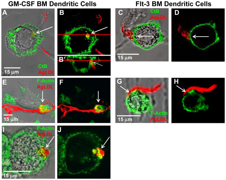
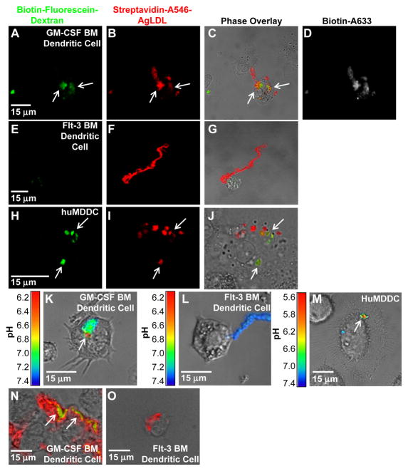
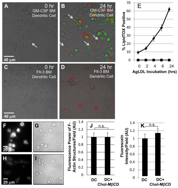
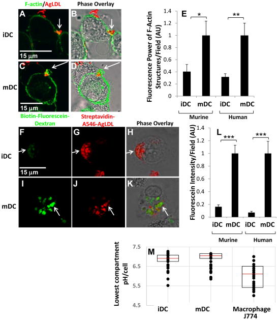
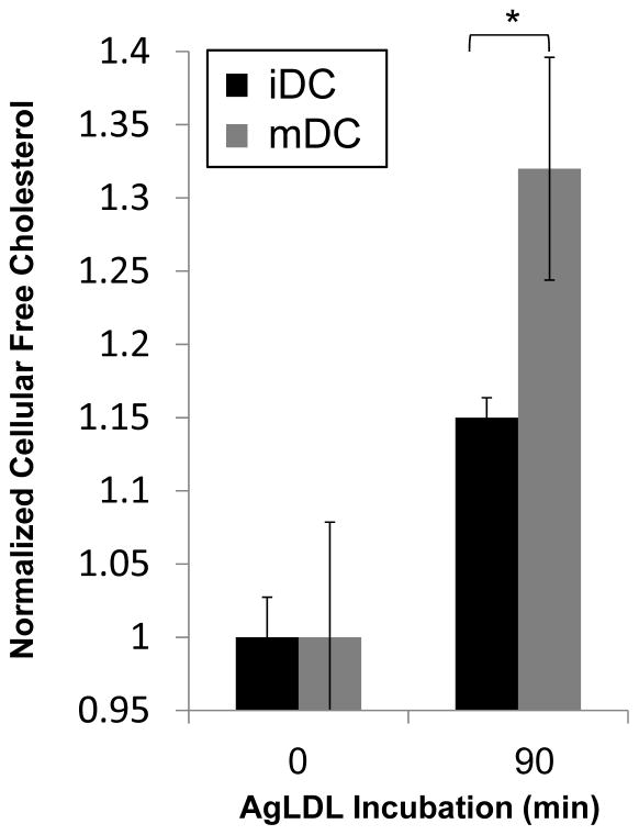
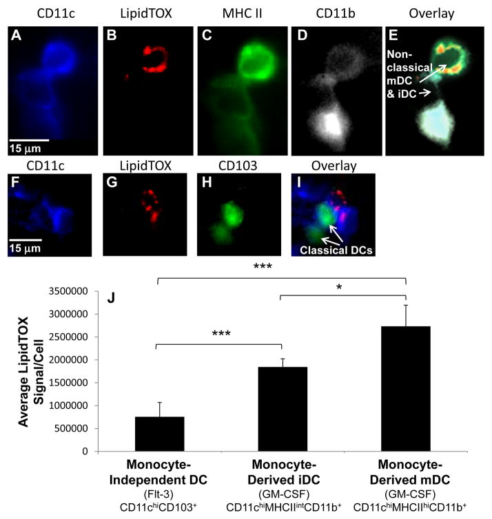
Approach and results: We demonstrate that both murine and human monocyte-derived dendritic cells use exophagy to degrade aggregated LDL, leading to foam cell formation, whereas monocyte-independent dendritic cells are unable to clear LDL aggregates by this mechanism. Exophagy is a catabolic process in which objects that cannot be internalized by phagocytosis (because of their size or association with extracellular structures) are initially digested in an extracellular acidic lytic compartment. Surprisingly, we found that monocyte-derived dendritic cells upregulate exophagy on maturation. This contrasts various forms of endocytic internalization in dendritic cells, which decrease on maturation. Finally, we show that our in vitro results are consistent with dendritic cell lipid accumulation in plaques of an ApoE(-/-) mouse model of atherosclerosis.

Conclusions: Our results show that monocyte-derived dendritic cells use exophagy to degrade aggregated LDL and become foam cells, whereas monocyte-independent dendritic cells are unable to clear LDL deposits. Furthermore, we find that exophagy is upregulated on dendritic cell maturation. Thus, exophagy-mediated foam cell formation in monocyte-derived dendritic cells could play a significant role in atherogenesis.

Keywords: LDL; atherosclerosis; dendritic cells; foam cells; lipoproteins; phagocytosis.


Figures

Figure 1
Figure 1. GM-CSF bone marrow-derived DCs form extracellular compartments at sites of contact with agLDL but flt-3 bone marrow-derived DCs do not
(A–J) GM-CSF BM (A, B and E, F and I, J) and flt-3 BM (C, D and G, H) DCs were incubated with Alexa546-agLDL (red) for 60 min and the interface between the aggregate and cells examined. (A–D) The plasma membrane was labeled via incubation with Alexa488-CtB (green) on ice for 3 min. Cells were than washed and fixed. Sites of contact between mature GM-CSF BM DCs and agLDL were labeled by CtB (arrows, A and B). An axial slice through the confocal stack at the position of the red line shows that, although the aggregate resembles a separate vesicle in the xy plane (B), it is contained in a compartment contiguous with the extracellular space (arrow, B′). Flt-3 BM DCs do not surround the aggregate with CtB positive membrane at sites of contact (arrows, C and D). (E–J) Cells were stained with Alexa488-phalloidin to show F-actin (green). An enrichment of F-actin was detected near the sites of contact with agLDL in GM-CSF BM (arrows, E and F) but not flt-3 BM (arrows, G and H) DCs. (I and J) Actin polymerization, at sites of contact with agLDL was also seen in mature huMDDCs (arrows, I and J).
Figure 2
Figure 2. GM-CSF BM DCs use exophagy to degrade agLDL but flt-3 BM DCs do not
(A–J) GM-CSF BM and flt-3 BM DC lysosomes were loaded with biotin-fluorescein-dextran via overnight incubation. DCs were subsequently incubated with streptavidin and Alexa546 dual-labeled agLDL for 90 min, fixed and permeabilized. Colocalization of dextran (green) with the aggregate (red) indicates areas of exocytosis in mature GM-CSF BM DCs (arrows, A–C). A short pulse of Alexa633-biotin prior to fixation confirmed that the agLDL was still contained in an extracellular compartment (D). No lysosome exocytosis to areas of contact with the aggregate was observed in flt-3 BM DCs (E–G). Lysosome exocytosis to an extracellular agLDL containing compartment was confirmed in mature huMDDCs (arrows, H–J). (K–M) GM-CSF BM and flt-3 BM DCs were incubated with CypHer 5E, a pH sensitive fluorophore, and Alexa488, a pH insensitive fluorophore, dual-labeled agLDL and the pH surrounding the aggregate was measured. When mature GM-CSF BM DCs interacted with the dual-labeled agLDL, regions of low pH could be seen at the contact sites (K). No acidification was observed in aggregates in contact with classical DCs (L). Regions of low pH were also seen in contact sites between mature huMDDCs and agLDL (M). (N and O) GM-CSF BM and flt-3 BM DCs were incubated with Alexa546-agLDL for 1 hr, fixed and stained with filipin to indicate free cholesterol. (N) Cholesterol accumulation can be seen in compartments containing agLDL in an immature GM-CSF BM DC (arrows). (O) No cholesterol generation was observed when flt-3 BM DCs were incubated with agLDL.
Figure 3
Figure 3. GM-CSF BM DCs incubated with agLDL form foam cells but flt-3 BM DCs do not
Immature GM-CSF BM DCs and flt-3 BM DCs were incubated with Alexa546-agLDL (red) for 0, 2, 4, 6 and 24 hrs. Cells were then fixed and stained with LipidTOX (green) to assess neutral lipid droplet formation. (A) Few LipidTOX positive non-classical DCs (arrow) are seen in the absence of agLDL. (B) GM-CSF BM DCs incubated with Alexa546-agLDL for 24 hrs form foam cells. Arrows indicate DCs in contact with agLDL containing LipidTOX positive droplets. (C) No LipidTOX positive flt-3 BM DCs are seen in the absence of agLDL. (D) Flt-3 BM DCs incubated with Alexa546-agLDL for 24hrs are negative for LipidTOX staining and do not form foam cells. (E) Quantification of the number of cells containing LipidTOX positive droplets as a function of agLDL incubation time. Diamonds: GM-CSF BM DCs, squares: flt-3 BM DCs. Error bars represent the standard error of the mean (sem). Data are pooled from 3 independent experiments for GM-CSF BM DCs and 2 independent experiments for flt-3 BM DCs. (F and G) Filipin and DIC image of immature GM-CSF BM DCs treated with 5mM cholesterol-MβCD for 15 min. (H and I) Filipin and DIC image of resting immature GM-CSF BM DCs. (J) Cholesterol loaded and resting immature GM-CSF BM DCs were incubated with Alexa546-agLDL for 60 min in the presence of an ACAT inhibitor followed by fixation, labeling of F-actin with Alexa488-phalloidin and quantification of the average local F-actin intensity per cell. (K) Cholesterol loaded and resting immature GM-CSF BM DCs with lysosomes labeled with biotin-fluorescein-dextran were incubated with streptavidin-Alexa546-labeled agLDL for 90 min in the presence of an ACAT inhibitor, fixed and permeabilized. The amount of biotin-fluorescein-dextran exocytosed was quantified. Data are pooled from 2 independent experiments. Student’s t test n.s. not significant.
Figure 4
Figure 4. Exophagy is upregulated with DC maturation
(A–D) GM-CSF BM iDCs and mDCs were incubated with Alexa546-agLDL for 60 min followed by fixation and labeling of F-actin with Alexa488-phalloidin. After 60 min, an enrichment of F-actin was detected near the sites of contact with agLDL in both GM-CSF BM iDCs (A and B) and mDCs (C and D). (E) Quantification of the average local F-actin intensity per cell in both murine and human monocyte derived iDCs and mDCs. (F–H) GM-CSF BM iDCs and mDCs were incubated with biotin-fluorescein-dextran overnight to deliver it to lysosomes. Cells were then exposed to streptavidin-Alexa546-labeled agLDL for 90 min, fixed and permeabilized. Lysosome exocytosis was observed to aggregate containing compartments in both GM-CSF BM iDCs (F–H) and mDCs (I–K). (L) Quantification of the amount of biotin-fluorescein-dextran exocytosed in both murine and human monocyte derived iDC and mDC. (M) GM-CSF BM iDCs, mDCs and J774 macrophage-like cells were incubated with CypHer 5E, a pH sensitive fluorophore, and Alexa488, a pH insensitive fluorophore, dual-labeled agLDL and the pH surrounding the aggregate measured. Quantification of the lowest pH achieved in the compartment at a single timepoint in iDCs, mDCs and J774 macrophage-like cells. The central mark on each box is the median, while the edges of the box represent the 25th and 75th percentiles. Error bars (E and L) represent the sem. * p ≤ 0.05, ** p ≤ 0.01, *** p ≤ 0.001 student’s t test. Data are pooled from 3 independent experiments.
Figure 5
Figure 5. Mature DC accumulate more free cholesterol than immature DC upon incubation with agLDL
Immature and mature DCs were incubated with agLDL for 0, 30 or 90 min. Lipids were extracted and free cholesterol determined by GC-MS analysis. Values were normalized for extraction using β-sitosterol and for protein content. No significant difference was seen in the free cholesterol content of iDC and mDC at 30 min (data not shown). Error bars represent the sem. * p ≤ 0.05 student’s t test. Data pooled from 3 independent experiments.
Figure 6
Figure 6. Lipid accumulation in DCs isolated from murine atherosclerotic plaques parallels in vitro results
Cells present in atherosclerotic plaques of 5 ApoE−/− mice on a high fat diet for 12 weeks were isolated and stained with monoclonal antibodies, for dissecting DC subsets, and LipidTOX-Red, for quantifying neutral lipid content. Quantitative analysis was used to delineate DC subsets. (A–E) Examples of a CD11chiMHCIIintCD11b+ moncyte-derived iDC and a CD11chiMHCIIhiCD11b+ monocyte-derived mDC (arrows, E). Cells with high CD11b expression were excluded from analysis. (F–I) Examples of CD103+CD11c+ monocyte-independent DCs (arrows, I). (J) Quantification of the LipidTOX signal per cell for each DC subset. Error bars represent the sem. * p ≤ 0.05, *** p ≤ 0.001 Kruskal-Wallis test followed by Wilcoxon rank-sum test.

References

    1. Bobryshev YV, Lord RS. Ultrastructural recognition of cells with dendritic cell morphology in human aortic intima. Contacting interactions of Vascular Dendritic Cells in athero-resistant and athero-prone areas of the normal aorta. Arch Histol Cytol. 1995;58:307–322. - PubMed
    1. Bobryshev YV, Lord RS. Structural heterogeneity and contacting interactions of vascular dendritic cells in early atherosclerotic lesions of the human aorta. J Submicrosc Cytol Pathol. 1996;28:49–60. - PubMed
    1. Bobryshev YV. Dendritic cells and their involvement in atherosclerosis. Current Opinion in Lipidology. 2000;11:511–517. - PubMed
    1. Bobryshev YV, Lord RS. S-100 positive cells in human arterial intima and in atherosclerotic lesions. Cardiovascular Research. 1995;29:689–696. - PubMed
    1. Bobryshev YV, Ikezawa T, Watanabe T. Formation of Birbeck granule-like structures in vascular dendritic cells in human atherosclerotic aorta. Lag-antibody to epidermal Langerhans cells recognizes cells in the aortic wall. Atherosclerosis. 1997;133:193–202. - PubMed

Publication types

MeSH terms

Substances