

Injectable shear-thinning hydrogels used to deliver endothelial progenitor cells, enhance cell engraftment, and improve ischemic myocardium
- PMID: 26293548
- PMCID: PMC4637242
- DOI: 10.1016/j.jtcvs.2015.07.035
Injectable shear-thinning hydrogels used to deliver endothelial progenitor cells, enhance cell engraftment, and improve ischemic myocardium
Abstract
Objectives: The clinical translation of cell-based therapies for ischemic heart disease has been limited because of low cell retention (<1%) within, and poor targeting to, ischemic myocardium. To address these issues, we developed an injectable hyaluronic acid (HA) shear-thinning hydrogel (STG) and endothelial progenitor cell (EPC) construct (STG-EPC). The STG assembles as a result of interactions of adamantine- and β-cyclodextrin-modified HA. It is shear-thinning to permit delivery via a syringe, and self-heals upon injection within the ischemic myocardium. This directed therapy to the ischemic myocardial border zone enables direct cell delivery to address adverse remodeling after myocardial infarction. We hypothesize that this system will enhance vasculogenesis to improve myocardial stabilization in the context of a clinically translatable therapy.
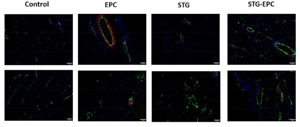
Methods: Endothelial progenitor cells (DiLDL(+) VEGFR2(+) CD34(+)) were harvested from adult male rats, cultured, and suspended in the STG. In vitro viability was quantified using a live-dead stain of EPCs. The STG-EPC constructs were injected at the border zone of ischemic rat myocardium after acute myocardial infarction (left anterior descending coronary artery ligation). The migration of the enhanced green fluorescent proteins from the construct to ischemic myocardium was analyzed using fluorescent microscopy. Vasculogenesis, myocardial remodeling, and hemodynamic function were analyzed in 4 groups: control (phosphate buffered saline injection); intramyocardial injection of EPCs alone; injection of the STG alone; and treatment with the STG-EPC construct. Hemodynamics and ventricular geometry were quantified using echocardiography and Doppler flow analysis.
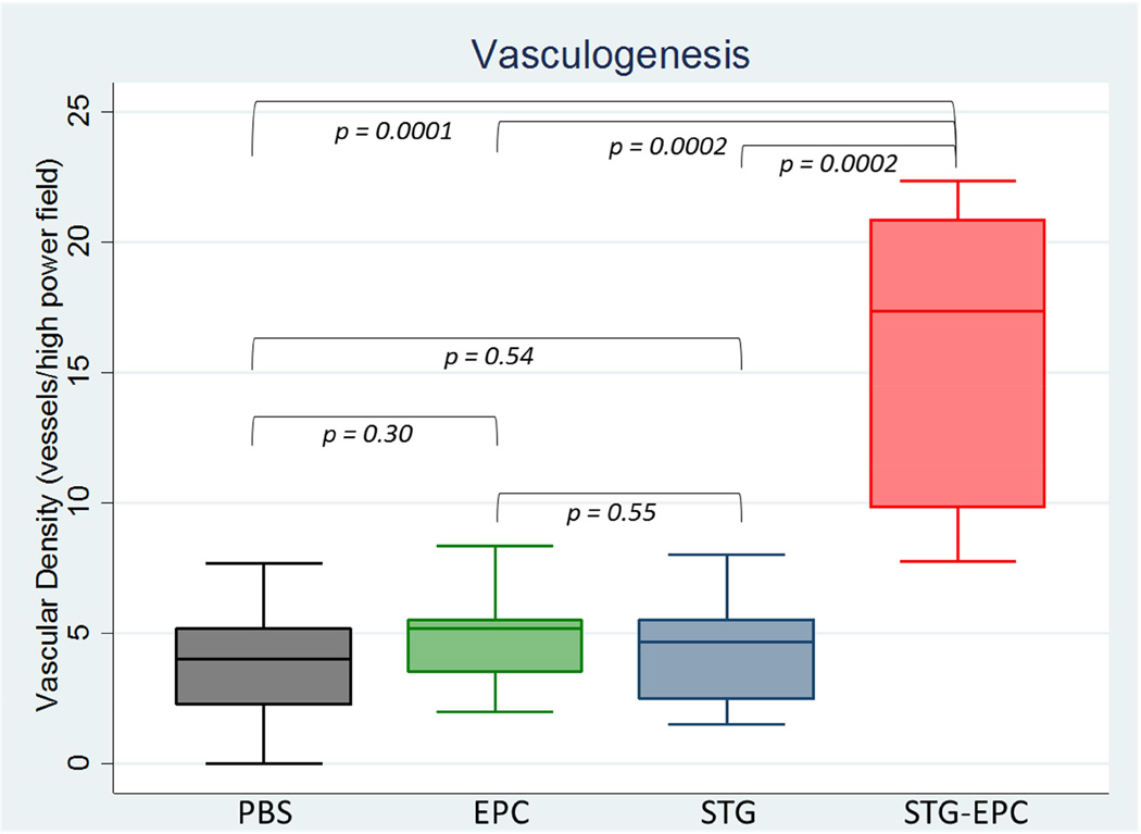
Results: Endothelial progenitor cells demonstrated viability within the STG. A marked increase in EPC engraftment was observed 1-week postinjection within the treated myocardium with gel delivery, compared with EPC injection alone (17.2 ± 0.8 cells per high power field (HPF) vs 3.5 cells ± 1.3 cells per HPF, P = .0002). A statistically significant increase in vasculogenesis was noted with the STG-EPC construct (15.3 ± 5.8 vessels per HPF), compared with the control (P < .0001), EPC (P < .0001), and STG (P < .0001) groups. Statistically significant improvements in ventricular function, scar fraction, and geometry were noted after STG-EPC treatment compared with the control.
Conclusions: A novel injectable shear-thinning HA hydrogel seeded with EPCs enhanced cell retention and vasculogenesis after delivery to ischemic myocardium. This therapy limited adverse myocardial remodeling while preserving contractility.
Keywords: cellular therapy; hydrogel; ischemic myocardium; shear-thinning gel.
Copyright © 2015 The American Association for Thoracic Surgery. Published by Elsevier Inc. All rights reserved.
Conflict of interest statement
Figures


Comment in
-
Discussion.J Thorac Cardiovasc Surg. 2015 Nov;150(5):1276-7. doi: 10.1016/j.jtcvs.2015.07.038. Epub 2015 Aug 17. J Thorac Cardiovasc Surg. 2015. PMID: 26293549 No abstract available.
-
Thankfully, back to basics.J Thorac Cardiovasc Surg. 2015 Nov;150(5):1278-9. doi: 10.1016/j.jtcvs.2015.07.081. Epub 2015 Jul 29. J Thorac Cardiovasc Surg. 2015. PMID: 26349595 No abstract available.
-
Stem cell therapy for heart failure: Out with the new and in with the old?J Thorac Cardiovasc Surg. 2015 Nov;150(5):1035-7. doi: 10.1016/j.jtcvs.2015.09.035. Epub 2015 Sep 16. J Thorac Cardiovasc Surg. 2015. PMID: 26546197 No abstract available.
References
-
- Araszkiewicz A, Grajek S, Lesiak M, Prech M, Pyda M, Janus M, et al. Effect of impaired myocardial reperfusion on left ventricular remodeling in patients with anterior wall acute myocardial infarction treated with primary coronary intervention. Am J Cardiol. 2006;98(6):725–728. - PubMed
-
- Bolognese L, Carrabba N, Parodi G, Santoro GM, Buonamici P, Cerisano G, et al. Impact of microvascular dysfunction on left ventricular remodeling and long-term clinical outcome after primary coronary angioplasty for acute myocardial infarction. Circulation. 2004;109(9):1121–1126. - PubMed
Publication types
MeSH terms
Substances
Grants and funding
LinkOut - more resources
Full Text Sources
Other Literature Sources
