

Segmentation of the lateral femoral notch sign with MRI using a new measurement technique
- PMID: 26293660
- PMCID: PMC4546148
- DOI: 10.1186/s12891-015-0677-0
Segmentation of the lateral femoral notch sign with MRI using a new measurement technique
Abstract
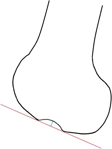
Background: The goal of this present study was to precisely determine the dimension and location of the impaction fracture on the lateral femoral condyle in patients with an ACL rupture.
Methods: All patients with post-injury bi-plane radiographs and MRI images after sustaining a tear to the anterior cruciate ligament were included. Lateral radiographs of the affected knee were inspected for a lateral femoral notch sign. MRIs of patients with a lateral condylopatellar sulcus ≥1.5 mm were used to segment and measure the lateral condylopatellar sulcus. The MRI examination was interpreted by an expert in musculoskeletal radiology. The study was approved by the ethics committee of the state of Salzburg.
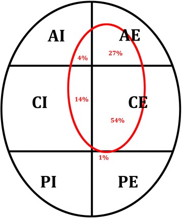
Results: A "lateral femoral notch sign"was seen in 50 patients. The average total surface area of the lateral femoral condyle was 3271.7 mm(2) (SD 739.5 mm(2)). The defect had a mean surface area of 266.1 mm(2) (SD 125.5 mm(2)), a mean volume of 456.5 mm(3) (SD 278.5 mm(3)), a mean depth of 3.0 mm (SD 0.8 mm). On average 169 mm(2) (SD 99.6 mm(2)) of the surface of the condyle were affected by the impaction fracture which corresponds to 5.2% (SD 2.8%) of the surface of the lateral femoral condyle. In 51 % the impaction fracture was located in the central-external area of the femoral condyle.
Conclusions: In cases of a clinically suspected ACL rupture lateral radiographs of the knee should be checked for a lateral femoral notch sign further MRI for confirmation should be performed. Knowing of the precise defect on the lateral femoral condyle is an additionally valuable information, as concomitant injuries to a rupture of the anterior cruciate ligament increase the risk for early-onset osteoarthritis in the future.
Figures

References
-
- Spindler KP, Schils JP, Bergfeld JA, Andrish JT, Weiker GG, Anderson TE, Piraino DW, Richmond BJ, Medendorp SV. Prospective study of osseous, articular, and meniscal lesions in recent anterior cruciate ligament tears by magnetic resonance imaging and arthroscopy. Am J Sports Med. 1993;21(4):551–557. doi: 10.1177/036354659302100412. - DOI - PubMed
-
- Murphy BJ, Smith RL, Uribe JW, Janecki CJ, Hechtman KS, Mangasarian RA. Bone signal abnormalities in the posterolateral tibia and lateral femoral condyle in complete tears of the anterior cruciate ligament: a specific sign? Radiology. 1992;182(1):221–224. doi: 10.1148/radiology.182.1.1727286. - DOI - PubMed
MeSH terms
LinkOut - more resources
Full Text Sources
Other Literature Sources
Medical
